. 2010 Feb 5;285(6):3554-3560.
doi: 10.1074/jbc.M109.063354. Epub 2009 Dec 12.

IIp45 inhibits cell migration through inhibition of HDAC6

Affiliations

IIp45 inhibits cell migration through inhibition of HDAC6

Ying Wu et al. J Biol Chem. .

Abstract

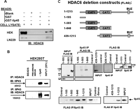
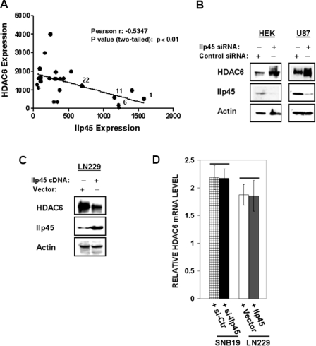
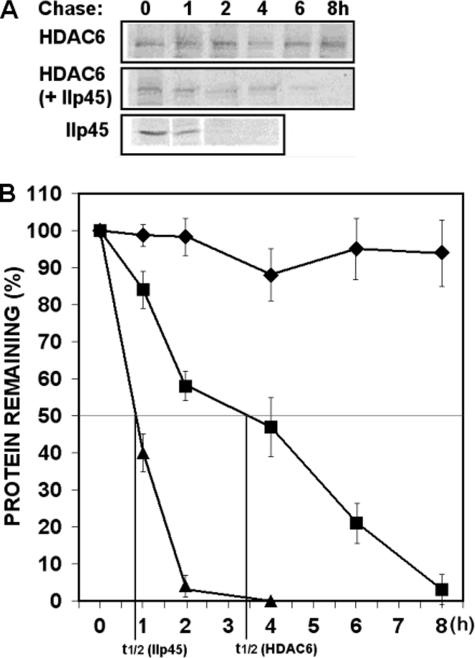
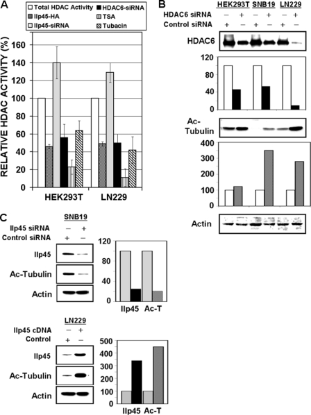
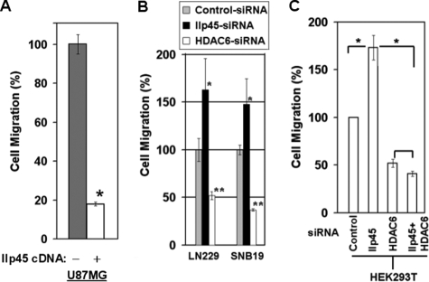
IIp45 (aka MIIP) is a newly discovered gene whose protein product inhibits cell migration. HDAC6 is a class IIb deacetylase that specifically deacetylates alpha-tubulin, modulates microtubule dynamics, and promotes cell migration. A yeast two-hybrid assay using IIp45 as bait identified HDAC6 protein as a binding partner of IIp45. This physical interaction of the two functionally antagonistic proteins was confirmed by glutathione S-transferase pulldown assay and co-immunoprecipitation assay in human cells. Serial deletion constructs of HDAC6 were used to characterize the interaction of HDAC6 and IIp45, and this analysis found that the two catalytic domains of HDAC6 protein are required for IIp45 binding. We examined the protein expression patterns of IIp45 and HDAC6 in glioma tissues. Elevated protein levels of HDAC6 were found in high grade glioma samples, in contrast to the decreased protein expression of IIp45. The potential negative regulation of HDAC6 expression by IIp45 was confirmed in cell lines with altered IIp45 expression by constitutive overexpression or small interfering RNA knockdown. Protein turnover study revealed that overexpression of IIp45 significantly reduces the intracellular protein stability of endogenous HDAC6, indicating a possible mechanism for the negative regulation of HDAC6 by IIp45. Results from the HDAC activity assay demonstrated that overexpressed IIp45 effectively decreases HDAC6 activity, increases acetylated alpha-tubulin, and reduces cell migration. The increased cell migration resulting from siIIp45 knockdown was significantly reversed by co-transfection of siHDAC6. Thus, we report here for the first time a novel mechanism by which IIp45 inhibits cell motility through inhibition of HDAC6.


Figures

FIGURE 1.
FIGURE 1.
Interaction between IIp45 and HDAC6. A, GST pulldown assay. Cell lysates of LN229 cells and HDAC6-transfected HEK293T cells were subjected to GST pulldown assay with glutathione-Sepharose beads, GST, and GST-IIp45 fusion protein, followed by SDS/PAGE and Western blot analysis using anti-HDAC6 antibody. B, IIp45-HDAC6 co-IP in HEK293T cells using anti-IIp45 or anti-HDAC6 antibody with preimmune serum or blank protein G beads as negative control. Proteins bound to protein G beads were collected, washed, and subjected to SDS-PAGE/Western blot (IB) analysis. C, binding domain analyses of HDAC6 for interaction with IIp45 in HDAC6-FLAG construct-transfected HEK293T cells. Top panel, schematic description of the constructs of FLAG-tagged wild-type HDAC6 and four truncated HDAC6: HDAC6(1–105), from which two catalytic domains (CAT1 and CAT2) are deleted; HDAC6(1–503), which contains the CAT1 but not the CAT2 domain; HDAC6(1–840), which contains both CAT1 and CAT2; and HDAC6 (493–1215), which contains the CAT2 but not the CAT1 domain. Middle and bottom panels, reciprocal co-IP assay of deletion HDAC6 and endogenous IIp45 in HEK293T cells transfected with wild-type or each of the deletion constructs using monoclonal anti-FLAG immunoprecipitation (IP) and anti-IIp45 serum from rabbit. INPUT, immunoblot of steady level of HDAC6 or IIp45 in cell lysates (10–20% of the amount, as indicated, of the same cell lysate sample used for immunoprecipitation).
FIGURE 2.
FIGURE 2.
Inversed protein expression pattern of IIP45 and HDAC6 in gliomas and human cell lines. A, protein extracts of four low grade glioma tissue samples (gliomas 1, 6, 11, and 22) and 20 high grade gliomas (glioblastoma multiforme) tissue samples analyzed for HDAC6 and IIp45. The level of β-actin was measured as a loading control. The results were quantified with densitometry and normalized with β-actin. Statistic correlation analysis was performed using the GraphPad Prism 5 program. B and C, Western blot analysis of HDAC6 steady levels in HEK293T and U87MG cells transfected with siIIp45 (B) or with the IIp45 expression vector (C). D, real time RT-PCR analysis of the HDAC6 mRNA levels in IIp45 knockdown or overexpressed cells. There is no statistical difference.
FIGURE 3.
FIGURE 3.
IIp45 reduces protein stability of HDAC6. Intracellular protein stability of endogenous HDAC6 was measured by protein turnover assay in IIp45 stable-transfected LN229 cells and control cell line. Cells were split and grown overnight to 50% confluence and radiolabeled with [35S]methionine in a pulse-chase experiment. Cell lysates were collected at time points 0, 1, 2, 4, 6, and 8 h of chase. Clarified cell lysates were used for immunoprecipitation of endogenous HDAC6 or overexpressed IIp45 in parallel samples. Immunoprecipitated proteins were resolved on SDS-PAGE and detected by fluorography. A, increased protein turnover rate of endogenous HDAC6 was shown in IIp45 stable-transfected cell line. B, graphic data of HDAC6 protein turnover was based on quantification of gel densitometry using the National Institutes of Health IMAGE program. Protein degradation curves from the top: endogenous HDAC6 in LN229 (diamonds), endogenous HDAC6 in LN229-IIp45-stable cell line (squares), IIp45 in LN229-IIp45-stable cell line (triangles).
FIGURE 4.
FIGURE 4.
IIp45 inhibits HDAC6 activity and increases intracellular acetylated α-tubulin level. A, HDAC activity assay was performed on cell lysates (100 μg of protein) of HEK293T and LN229 cells. Cells were transfected with wild-type IIp45-HA or treated by IIp45-siRNA or HDAC6-siRNA. Data were calculated with control reagent-treated cells and are presented as a percentage of HDAC activity compared with the total HDAC activity (as 100%) in untreated parental cells. Bars in each cell line from left: untreated parental cells HDAC activity (as 100%; white bars), percentage of the HDAC activity in IIp45-transiently transfected cells compared with parental (dark gray bars), percentage of the HDAC activity in siIIp45-treated cells (light gray bars), percentage of HDAC activity in siHDAC6-treated cells (black bars), percentage of HDAC activity in parental cell lysates measured by HDAC activity assays containing HDAC inhibitors TSA (0.5 μm) (striped bars) and tubacin (1 μm) (hatched bars). Error bars, mean ± S.E. B and C, Western blotting of acetylated α-tubulin. The intensity of the bands was quantified with densitometry and normalized with that of β-actin loading control. Values are presented as bar graphs in percentage relative to control (100%). B, increased level of acetylated α-tubulin in HDAC6 siRNA (40 nm) transfected HEK293T, SNB19, and LN229. C, effects of altered IIp45 expression on acetylated α-tubulin, showing a decreased level in siIIp45-treated SNB19 (upper panels, compared with control siRNA) and increased level in IIp45-transfected LN229 (lower panels, compared with control vector).
FIGURE 5.
FIGURE 5.
IIp45 inhibits cell migration through inhibition of HDAC6. Semiconfluent cells were split into triplicate wells with a pore size of 8 μm. The chambers were incubated at 37 °C in a 5% CO2 incubator for 3 h (U87MG), 6 h (SNB19), or 15 h (for LN229 and HEK293T). Migrated cells were then fixed, stained, and counted. Mean value ± S.E. (error bars) were calculated based on triplicate sample setting and repeated three times. A, IIp45 stable-transfected U87MG cell line compared with the parental cell line (*, p < 0.05). B, siIIp45 (20 nm) and siHDAC6 (40 nm) knockdown showed opposite effects on cell migration in both LN229 (left) and SNB19 (right) cell lines (compared with control siRNA, *, p < 0.05; **, p < 0.002, respectively). C, single and double siRNA knockdown in HEK293T cells (*, p < 0.05).

Similar articles

Cited by

References

    1. Wehrle-Haller B., Imhof B. A. (2003) Int. J. Biochem. Cell Biol. 35, 39–50 - PubMed
    1. Cunha-Ferreira I., Bento I., Bettencourt-Dias M. (2009) Traffic 10, 482–498 - PubMed
    1. Takemura R., Okabe S., Umeyama T., Kanai Y., Cowan N. J., Hirokawa N. (1992) J. Cell Sci. 103, 953–964 - PubMed
    1. Hammond J. W., Cai D., Verhey K. J. (2008) Curr. Opin. Cell Biol. 20, 71–76 - PMC - PubMed
    1. De Ruijter A. J. M., van Gennip A. H., Caron H. N., Kemp S., van Kuilenburgi A. B. P. (2003) Biochem. J. 370, 737–749 - PMC - PubMed

Publication types

MeSH terms

LinkOut - more resources