

Mechanisms of epigenetic silencing of the Rassf1a gene during estrogen-induced breast carcinogenesis in ACI rats
- PMID: 20008439
- PMCID: PMC2899845
- DOI: 10.1093/carcin/bgp304
Mechanisms of epigenetic silencing of the Rassf1a gene during estrogen-induced breast carcinogenesis in ACI rats
Abstract
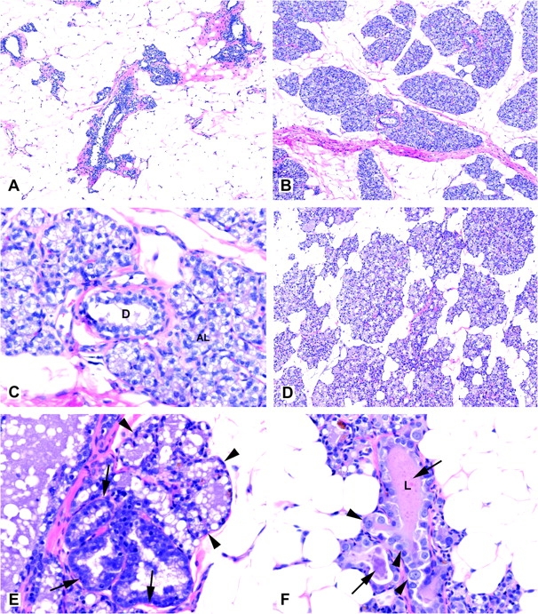
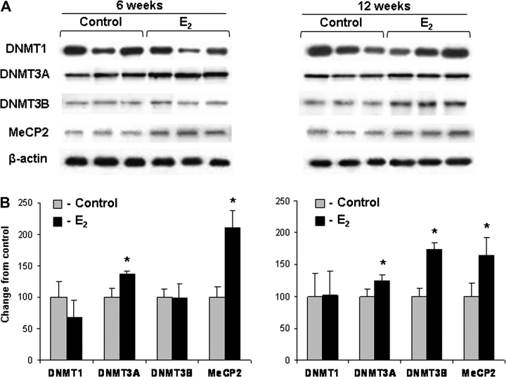
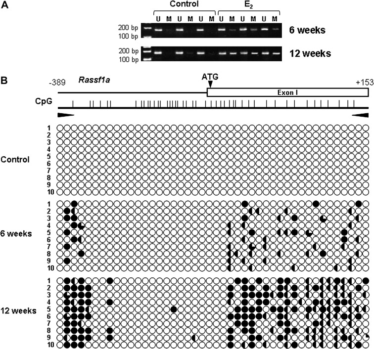
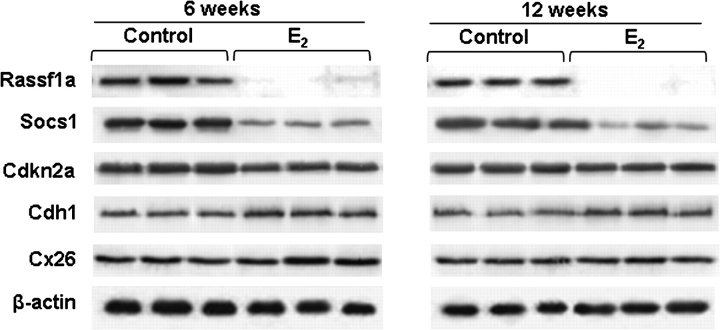
Breast cancer, the most common malignancy in women, emerges through a multistep process, encompassing the progressive sequential evolution of morphologically distinct stages from a normal cell to hyperplasia (with and without atypia), carcinoma in situ, invasive carcinoma and metastasis. The success of treatment of breast cancer could be greatly improved by the detection at early stages of cancer. In the present study, we investigated the underlying molecular mechanisms involved in breast carcinogenesis in Augustus and Copenhagen-Irish female rats, a cross between the ACI strains, induced by continuous exposure to 17beta-estradiol. The results of our study demonstrate that early stages of estrogen-induced breast carcinogenesis are characterized by altered global DNA methylation, aberrant expression of proteins responsible for the proper maintenance of DNA methylation pattern and epigenetic silencing of the critical Rassf1a (Ras-association domain family 1, isoform A) tumor suppressor gene. Interestingly, transcriptional repression of the Rassf1a gene in mammary glands during early stages of breast carcinogenesis was associated with an increase in trimethylation of histones H3 lysine 9 and H3 lysine 27 and de novo CpG island methylation and at the Rassf1a promoter and first exon. In conclusion, we demonstrate that epigenetic alterations precede formation of preneoplastic lesions indicating the significance of epigenetic events in induction of oncogenic pathways in early stages of carcinogenesis.
Figures
Similar articles
-
Estrogen-induced rat breast carcinogenesis is characterized by alterations in DNA methylation, histone modifications and aberrant microRNA expression.Cell Cycle. 2007 Aug 15;6(16):2010-8. doi: 10.4161/cc.6.16.4549. Epub 2007 Jun 6. Cell Cycle. 2007. PMID: 17700064
-
Chromatin inactivation precedes de novo DNA methylation during the progressive epigenetic silencing of the RASSF1A promoter.Mol Cell Biol. 2005 May;25(10):3923-33. doi: 10.1128/MCB.25.10.3923-3933.2005. Mol Cell Biol. 2005. PMID: 15870267 Free PMC article.
-
Frequent silencing of RASSF1A via promoter methylation in follicular thyroid hyperplasia: a potential early epigenetic susceptibility event in thyroid carcinogenesis.JAMA Surg. 2014 Nov;149(11):1146-52. doi: 10.1001/jamasurg.2014.1694. JAMA Surg. 2014. PMID: 25229773
-
Gene methylation in gastric cancer.Clin Chim Acta. 2013 Sep 23;424:53-65. doi: 10.1016/j.cca.2013.05.002. Epub 2013 May 10. Clin Chim Acta. 2013. PMID: 23669186 Review.
-
The tumor suppressor RASSF1A in human carcinogenesis: an update.Histol Histopathol. 2005 Apr;20(2):645-63. doi: 10.14670/HH-20.645. Histol Histopathol. 2005. PMID: 15736067 Review.
Cited by
-
Oral Contraceptive Use May Modulate Global Genomic DNA Methylation and Promoter Methylation of APC1 and ESR1.Asian Pac J Cancer Prev. 2017 Sep 27;18(9):2361-2366. doi: 10.22034/APJCP.2017.18.9.2361. Asian Pac J Cancer Prev. 2017. PMID: 28950679 Free PMC article.
-
Epigenetic regulation of estrogen signaling in breast cancer.Epigenetics. 2013 Mar;8(3):237-45. doi: 10.4161/epi.23790. Epub 2013 Jan 30. Epigenetics. 2013. PMID: 23364277 Free PMC article. Review.
-
Body mass and DNA promoter methylation in breast tumors in the Western New York Exposures and Breast Cancer Study.Am J Clin Nutr. 2011 Sep;94(3):831-8. doi: 10.3945/ajcn.110.009365. Epub 2011 Jul 20. Am J Clin Nutr. 2011. PMID: 21775555 Free PMC article.
-
MicroRNA-mediated drug resistance in breast cancer.Clin Epigenetics. 2011 Aug;2(2):171-185. doi: 10.1007/s13148-011-0040-8. Epub 2011 Jun 27. Clin Epigenetics. 2011. PMID: 21949547 Free PMC article.
-
Maternal weight during pregnancy and risk of childhood acute lymphoblastic leukemia in offspring.Leukemia. 2025 Mar;39(3):590-598. doi: 10.1038/s41375-025-02517-6. Epub 2025 Jan 26. Leukemia. 2025. PMID: 39865137 Free PMC article.
References
-
- Glass AG, et al. Breast cancer incidence, 1980–2006: combined roles of menopausal hormone therapy, screening mammography, and estrogen receptor status. J. Natl Cancer Inst. 2007;99:1152–1161. - PubMed
-
- Jemal A, et al. Cancer statistics, 2008. CA Cancer J. Clin. 2008;58:71–96. - PubMed
-
- Jemal A, et al. Cancer statistics, 2009. CA Cancer J. Clin. 2009;59:225–249. - PubMed
-
- Hanahan D, et al. The hallmarks of cancer. Cell. 2000;100:57–70. - PubMed
