. 2010 Feb 15;588(Pt 4):617-32.
doi: 10.1113/jphysiol.2009.182147. Epub 2009 Dec 14.

Electrophysiological identification of glucose-sensing neurons in rat nodose ganglia

Affiliations

Electrophysiological identification of glucose-sensing neurons in rat nodose ganglia

Gintautas Grabauskas et al. J Physiol. .

Abstract

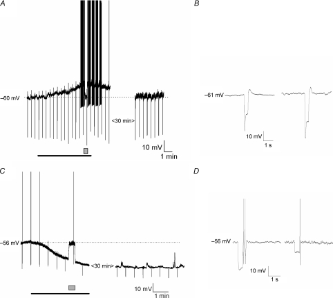
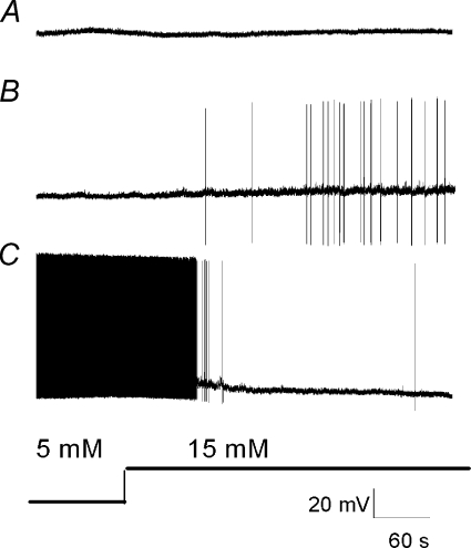
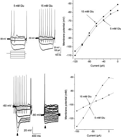
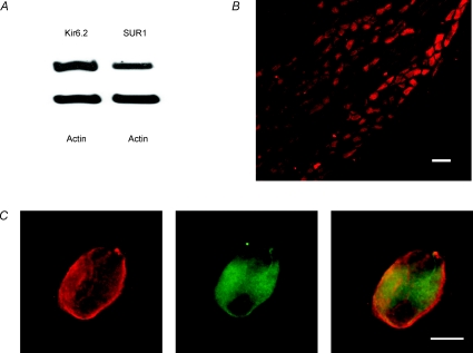
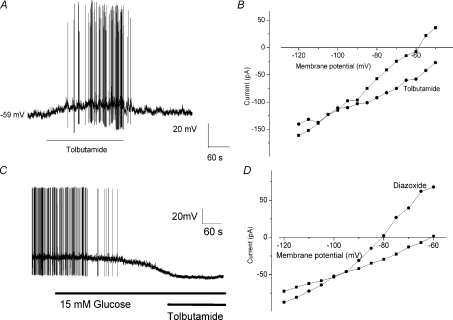
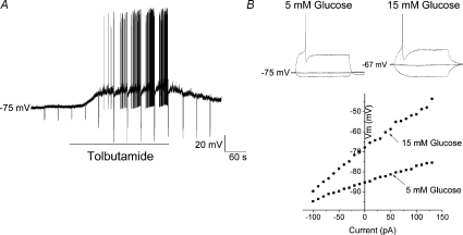
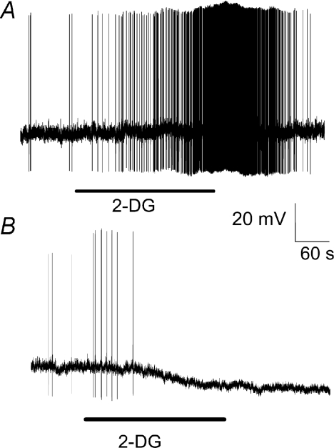
The vagal afferent system is strategically positioned to mediate rapid changes in motility and satiety in response to systemic glucose levels. In the present study we aimed to identify glucose-excited and glucose-inhibited neurons in nodose ganglia and characterize their glucose-sensing properties. Whole-cell patch-clamp recordings in vagal afferent neurons isolated from rat nodose ganglia demonstrated that 31/118 (26%) neurons were depolarized after increasing extracellular glucose from 5 to 15 mm; 19/118 (16%) were hyperpolarized, and 68/118 were non-responsive. A higher incidence of excitatory response to glucose occurred in gastric- than in portal vein-projecting neurons, the latter having a higher incidence of inhibitory response. In glucose-excited neurons, elevated glucose evoked membrane depolarization (11 mV) and an increase in membrane input resistance (361 to 437 M). Current reversed at 99 mV. In glucose-inhibited neurons, membrane hyperpolarization (13 mV) was associated with decreased membrane input resistance (383 to 293 M). Current reversed at 97 mV. Superfusion of tolbutamide, a K(ATP) channel sulfonylurea receptor blocker, elicited identical glucose-excitatory but not glucose-inhibitory responses. Kir6.2 shRNA transfection abolished glucose-excited but not glucose-inhibited responses. Phosphatidylinositol bisphosphate (PIP(2)) depletion using wortmannin increased the fraction of glucose-excited neurons from 26% to 80%. These results show that rat nodose ganglia have glucose-excited and glucose-inhibited neurons, differentially distributed among gastric- and portal vein-projecting nodose neurons. In glucose-excited neurons, glucose metabolism leads to K(ATP) channel closure, triggering membrane depolarization, whereas in glucose-inhibited neurons, the inhibitory effect of elevated glucose is mediated by an ATP-independent K(+) channel. The results also show that PIP(2) can determine the excitability of glucose-excited neurons.


Figures

Figure 2
Figure 2. Effects of glucose on the membrane properties of nodose ganglia neurons
A, continues membrane potential recording shows that increase in extracellular glucose concentration from 5 to 15 mm (bar) depolarized the membrane potential sufficient to elicit action potentials. The depolarization was associated with the increase in neuronal input resistance; the neuronal input resistance was tested every 40 s by injecting 500 ms, 100 pA amplitude current pulses (negative membrane potential deflections). To evaluate changes in the membrane input resistance the membrane potential was current clamped back (box). Note that after glucose concentration was restored to 5 mm, membrane potential and input resistance returned to baseline within 30 min. B, the same experiment illustrated in A is depicted on an expanded time scale illustrating the membrane potential response to injection of 100 pA current pulse at 5 mm (left) and 15 mm (right) glucose concentration (potential was current clamped back). C, continues membrane potential recording shows that increase in extracellular glucose concentration from 5 mm to 15 mm hyperpolarized the membrane potential in a glucose inhibited neuron. The hyperpolarization was associated with a decrease in neuronal input resistance. To evaluate changes in the membrane input resistance the membrane potential was current clamped back (box). Note that the effect of hyperpolarization was sustained after 30 min of recording. D, the same experiment illustrated in C is depicted on an expanded time scale to show the membrane potential response to injection of 100 pA current pulse at 5 mm (left) and 15 mm (right) glucose concentration (potential was current clamped back).
Figure 1
Figure 1
Continuous membrane potential recordings depict typical responses of glucose-insensitive (A), glucose-excited (B) and glucose-inhibited (C) nodose ganglia neurons to an increase in extracellular glucose (5 to 15 mm)
Figure 3
Figure 3. Glucose levels modulate the membrane properties of nodose ganglia neurons
A, current–clamp responses show that increase in extracellular glucose (Glu) increased the excitability of glucose-excited neurons, and this was associated with an increase in membrane input resistance; note the increase in amplitude of the negative membrane potential deflection evoked by negative current pulses. B, current–voltage (I–V) relationships from a glucose-excited neuron show that an increase in extracellular glucose concentration increased the slope of the relationship; the effect reversed at −108 mV, close to the estimated K+ equilibrium potential. C, current-clamp responses demonstrate that an increase in extracellular glucose hyperpolarized the recorded neuron from −60 mV to −80 mV. This increase also decreased membrane input resistance and suppressed the action potentials generated by positive current pulses. The arrowheads indicate the time when the data for I–V relationship were acquired. D, current–voltage relationship for a glucose-inhibited type of neuron demonstrates that an increase of glucose concentration reduced the slope of the relationship; the effect reversed at −98 mV, close to the estimated K+ equilibrium potential.
Figure 4
Figure 4. Kir6.2 and SUR1 protein expression in the rat nodose ganglia
A, representative immunoblot from the nodose ganglia shows Kir6.2 and SUR1 protein expression; actin used as loading control. B, immunohistochemistry studies to identify neurons containing the inwardly rectifying K+ channels in nodose ganglion cross-section. Calibration bar, 100 μm. C, representative immunofluorescence staining for SUR1 (left), the inwardly rectifying K+ channel Kir6.2 (middle), and overlay (right) in a nodose ganglion neuron. Calibration bar, 20 μm.
Figure 5
Figure 5. Effects of KATP channel modulators on membrane properties of nodose ganglia neurons
A, continuous membrane potential recordings depict the typical response to extracellular superfusion of tolbutamide (200 μm) in an isolated nodose ganglion neuron. B, tolbutamide (200 μm) shifted the I–V relationship in a depolarized direction, and changed the slope of the relationship, indicating increased input resistance. I–V relationships crossed at −90 mV. The I–V relationship was recorded in the presence of 2 mm extracellular glucose. C, continuous membrane potential recording demonstrating that tolbutamide (200 μm) did not attenuate the duration or the amplitude of the hyperpolarization current generated by ‘high’ glucose concentration, indicating that KATP channels do not modulate the excitability of glucose-inhibited neurons. D, extracellular application of diazoxide (100 μm), a KATP channel activator, generated a shift of the I–V relationship, hyperpolarizing duration and decreasing membrane input resistance. The I–V relationship was recorded in the presence of 15 mm extracellular glucose.
Figure 6
Figure 6. Lentivirus-based transfection of shRNA for Kir6.2 suppresses endogenous protein and mRNA expression
A, Western blot demonstrating a significant reduction of Kir6.2 protein 96 h after the primary nodose ganglion neuron cultures were transfected with lenti-Kir6.2 shRNA. Actin was used as a loading control. B, photomicrograph of nodose ganglion neuron transfected with lenti-shKir6.2 and cultured for 96 h displaying intense EGFP signal (arrow). C, faint immunostain signal for anti-Kir6.2 antibody in the same neuron shown in B (arrow). D, photomicrograph of nodose ganglion neuron transfected with scrambled siRNA and cultured for 96 h displaying intense EGFP signal (arrow). E, immunostain signal for anti-Kir6.2 antibody in the same neuron shown in D (arrow).
Figure 7
Figure 7. Effects of wortmannin on the membrane properties of nodose ganglia neurons
A, continuous membrane potential recording from wortmannin (50 nm)-treated neuron in response to extracellular superfusion of tolbutamide (200 μm). Tolbutamide depolarized the recoded neuron from −75 to −46 mV, an effect associated with the increase in membrane input resistance from 110 to 596 MΩ. The effect of tolbutamide was reversible. B, wortmannin treatment enhanced the neuronal sensitivity to glucose. Current-clamp recordings demonstrate that an increase in extracellular glucose concentration depolarized the recorded neuron from −85 to −67 mV and increased the membrane input resistance from 98 to 250 MΩ. In addition, the glucose concentration transition decreased the threshold for action potential triggering from −560 to 150 pA. The I–V relationship demonstrates that the glucose effect reversed at ∼−110 mV, which is close to the theoretical K+ ion reversal potential in the conditions recorded.
Figure 8
Figure 8
Continuous membrane potential recording demonstrates that extracellular superfusion of 2-deoxyglucose (2-DG), a glucose metabolism inhibitor, generated either depolarization or hyperpolarization of neuronal membrane potential

Similar articles

Cited by

References

    1. Adachi A, Shimizu N, Oomura Y, Kobashi M. Convergence of hepatoportal glucose-sensitive afferent signals to glucose-sensitive units within the nucleus of the solitary tract. Neurosci Lett. 1984;46:215–218. - PubMed
    1. Balfour RH, Hansen AM, Trapp S. Neuronal responses to transient hypoglycaemia in the dorsal vagal complex of the rat brainstem. J Physiol. 2006;570:469–484. - PMC - PubMed
    1. Baukrowitz T, Schulte U, Oliver D, Herlitze S, Krauter T, Tucker SJ, Ruppersberg JP, Fakler B. PIP2 and PIP as determinants for ATP inhibition of KATP channels. Science. 1998;282:1141–1144. - PubMed
    1. Burdakov D, Jensen LT, Alexopoulos H, Williams RH, Fearon IM, O’Kelly I, Gerasimenko O, Fugger L, Verkhratsky A. Tandem-pore K+ channels mediate inhibition of orexin neurons by glucose. Neuron. 2006;50:711–722. - PubMed
    1. Burdakov D, Lesage F. Glucose-induced inhibition: how many ionic mechanisms? Acta Physiol (Oxf) 2009 in press. - PMC - PubMed

Publication types

MeSH terms

Associated data