

Cigarette smoke exposure impairs pulmonary bacterial clearance and alveolar macrophage complement-mediated phagocytosis of Streptococcus pneumoniae
- PMID: 20008540
- PMCID: PMC2825918
- DOI: 10.1128/IAI.00963-09
Cigarette smoke exposure impairs pulmonary bacterial clearance and alveolar macrophage complement-mediated phagocytosis of Streptococcus pneumoniae
Abstract
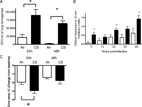
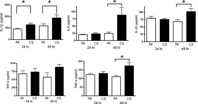
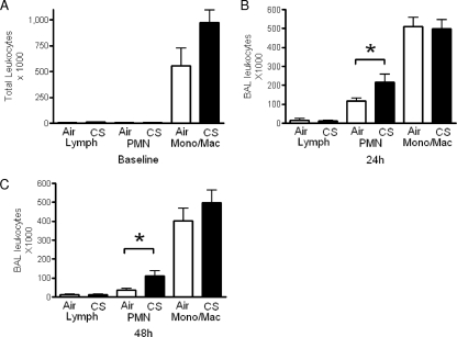
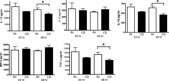
Cigarette smoke exposure increases the risk of pulmonary and invasive infections caused by Streptococcus pneumoniae, the most commonly isolated organism from patients with community-acquired pneumonia. Despite this association, the mechanisms by which cigarette smoke exposure diminishes host defense against S. pneumoniae infections are poorly understood. In this study, we compared the responses of BALB/c mice following an intratracheal challenge with S. pneumoniae after 5 weeks of exposure to room air or cigarette smoke in a whole-body exposure chamber in vivo and the effects of cigarette smoke on alveolar macrophage phagocytosis of S. pneumoniae in vitro. Bacterial burdens in cigarette smoke-exposed mice were increased at 24 and 48 h postinfection, and this was accompanied by a more pronounced clinical appearance of illness, hypothermia, and increased lung homogenate cytokines interleukin-1beta (IL-1beta), IL-6, IL-10, and tumor necrosis factor alpha (TNF-alpha). We also found greater numbers of neutrophils in bronchoalveolar lavage fluid recovered from cigarette smoke-exposed mice following a challenge with heat-killed S. pneumoniae. Interestingly, overnight culture of alveolar macrophages with 1% cigarette smoke extract, a level that did not affect alveolar macrophage viability, reduced complement-mediated phagocytosis of S. pneumoniae, while the ingestion of unopsonized bacteria or IgG-coated microspheres was not affected. This murine model provides robust additional support to the hypothesis that cigarette smoke exposure increases the risk of pneumococcal pneumonia and defines a novel cellular mechanism to help explain this immunosuppressive effect.
Figures

Similar articles
-
Trivalent pneumococcal protein recombinant vaccine protects against lethal Streptococcus pneumoniae pneumonia and correlates with phagocytosis by neutrophils during early pathogenesis.Vaccine. 2015 Feb 18;33(8):993-1000. doi: 10.1016/j.vaccine.2015.01.014. Epub 2015 Jan 15. Vaccine. 2015. PMID: 25597944
-
Exposure to cigarette smoke impairs alveolar macrophage functions during halothane and isoflurane anesthesia in rats.Anesthesiology. 1999 Dec;91(6):1823-33. doi: 10.1097/00000542-199912000-00037. Anesthesiology. 1999. PMID: 10598627
-
Smoke exposure and ethanol ingestion modulate intrapulmonary polymorphonuclear leukocyte killing, but not recruitment or phagocytosis.Alcohol Clin Exp Res. 2006 Sep;30(9):1599-607. doi: 10.1111/j.1530-0277.2006.00192.x. Alcohol Clin Exp Res. 2006. PMID: 16930223
-
Alveolar macrophages in pulmonary host defence the unrecognized role of apoptosis as a mechanism of intracellular bacterial killing.Clin Exp Immunol. 2013 Nov;174(2):193-202. doi: 10.1111/cei.12170. Clin Exp Immunol. 2013. PMID: 23841514 Free PMC article. Review.
-
Cigarette smoke exposure and alveolar macrophages: mechanisms for lung disease.Thorax. 2022 Jan;77(1):94-101. doi: 10.1136/thoraxjnl-2020-216296. Epub 2021 May 13. Thorax. 2022. PMID: 33986144 Free PMC article. Review.
Cited by
-
The Association of Toll-like Receptor-9 Gene Single-Nucleotide Polymorphism and AK155(IL-26) Serum Levels with Chronic Obstructive Pulmonary Disease Exacerbation Risk: A Case-Controlled Study with Bioinformatics Analysis.Biomedicines. 2025 Mar 3;13(3):613. doi: 10.3390/biomedicines13030613. Biomedicines. 2025. PMID: 40149591 Free PMC article.
-
Oxidative Inactivation of the Proteasome Augments Alveolar Macrophage Secretion of Vesicular SOCS3.Cells. 2020 Jun 30;9(7):1589. doi: 10.3390/cells9071589. Cells. 2020. PMID: 32630102 Free PMC article.
-
Glucocorticoid-Augmented Efferocytosis Inhibits Pulmonary Pneumococcal Clearance in Mice by Reducing Alveolar Macrophage Bactericidal Function.J Immunol. 2015 Jul 1;195(1):174-84. doi: 10.4049/jimmunol.1402217. Epub 2015 May 18. J Immunol. 2015. PMID: 25987742 Free PMC article.
-
E-Cigarette (E-Cig) Liquid Composition and Operational Voltage Define the In Vitro Toxicity of Δ8Tetrahydrocannabinol/Vitamin E Acetate (Δ8THC/VEA) E-Cig Aerosols.Toxicol Sci. 2022 May 26;187(2):279-297. doi: 10.1093/toxsci/kfac047. Toxicol Sci. 2022. PMID: 35478015 Free PMC article.
-
Cigarette smoke depletes alveolar macrophages and delays clearance of Legionella pneumophila.Am J Physiol Lung Cell Mol Physiol. 2023 Mar 1;324(3):L373-L384. doi: 10.1152/ajplung.00268.2022. Epub 2023 Jan 31. Am J Physiol Lung Cell Mol Physiol. 2023. PMID: 36719079 Free PMC article.
References
-
- Ali, F., M. E. Lee, F. Iannelli, G. Pozzi, T. J. Mitchell, R. C. Read, and D. H. Dockrell. 2003. Streptococcus pneumoniae-associated human macrophage apoptosis after bacterial internalization via complement and Fc-gamma receptors correlates with intracellular bacterial load. J. Infect. Dis. 188:1119-1131. - PubMed
-
- Almirall, J., I. Bolibar, M. Serra-Prat, J. Roig, I. Hospital, E. Carandell, M. Agusti, P. Ayuso, A. Estela, A. Torres, et al. 2008. New evidence of risk factors for community-acquired pneumonia: a population-based study. Eur. Respir. J. 31:1274-1284. - PubMed
-
- Arcavi, L., and N. Benowitz. 2004. Cigarette smoking and infection. Arch. Intern. Med. 164:2206-2216. - PubMed
Publication types
MeSH terms
Substances
Grants and funding
LinkOut - more resources
Full Text Sources
Molecular Biology Databases
