. 2010 Feb;30(4):985-94.
doi: 10.1128/MCB.00909-09. Epub 2009 Dec 14.

The ubiquitin ligase Hul5 promotes proteasomal processivity

Affiliations

The ubiquitin ligase Hul5 promotes proteasomal processivity

Sharon Aviram et al. Mol Cell Biol. 2010 Feb.

Abstract

The 26S proteasome is a large cytoplasmic protease that degrades polyubiquitinated proteins to short peptides in a processive manner. The proteasome 19S regulatory subcomplex tethers the target protein via its polyubiquitin adduct and unfolds the target polypeptide, which is then threaded into the proteolytic site-containing 20S subcomplex. Hul5 is a 19S subcomplex-associated ubiquitin ligase that elongates ubiquitin chains on proteasome-bound substrates. We isolated hul5 Delta as a mutation with which fusions of an unstable cyclin to stable reporter proteins accumulate as partially processed products. These products appear transiently in the wild type but are strongly stabilized in 19S ATPase mutants and in the hul5 Delta mutant, supporting a role for the ATPase subunits in the unfolding of proteasome substrates before insertion into the catalytic cavity and suggesting a role for Hul5 in the processive degradation of proteins that are stalled on the proteasome.


Figures

FIG. 1.
FIG. 1.
Expression of Ura3-Pcl5 in hul5Δ mutant cells generates a 36-kDa Ura3-containing species. (A) The schematic drawing of the fusion protein indicates the positions of the Ura3 and Pcl5 moieties, with the intervening HA tag indicated in dark gray. Cells were induced with CuSO4 and treated with cycloheximide (CHX) as detailed in Materials and Methods. Upper panel: detection of the Ura3-HA-Pcl5 fusion with the HA.11 antibody (α-HA). Lower panel: detection of the same membrane with an anti-Pcl5 antiserum (α-Pcl5). (B) Mass-spectrometry analysis of the 36-kDa species after digestion with trypsin (see Materials and Methods for details). The origins of the identified peptides are indicated, and their distribution on the Ura3-Pcl5 sequence is depicted schematically.
FIG. 2.
FIG. 2.
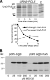
The Ura3-Pcl5 processing product is a consequence of proteasome activity. (A) Pulse-chase analysis of Ura3-Pcl5 degradation in hul5Δ mutant cells. Cells carrying plasmid KB1432 expressing the Ura3-Pcl5 fusion protein were labeled for 2 min and chased with cold methionine. C, cells carrying a vector plasmid. The 36-kDa processing product is indicated with an arrowhead. The graph depicts the quantitation of the experiment shown; the percentage of the processing product is indicated relative to the full-length protein at t0 of the chase, corrected for the smaller number of methionines and cysteines (the residues labeled with 35S) in the processing product, namely, 11, versus 20 in the full-length Ura3-Pcl5 fusion protein. (B) Accumulation of Ura3-Pcl5 and its processing product upon proteasome inhibition. Strains KY775 (pdr5 erg6) and KY1270 (pdr5 erg6 hul5) carrying plasmid KY1169 were incubated for 20 min with 0.2 mM CuSO4 (Ura3-Pcl5 induction) and with the indicated amounts of the proteasome inhibitor MG132 before protein extraction.
FIG. 3.
FIG. 3.
The Ura3-Pcl5 processing product is not inherently unstable. (A) Characterization of the Ura3-Pcl5 processing product size. Wild-type cells expressing Ura3 fused to various fragments of Pcl5 from plasmids KB1908 (1-46), KB1909 (1-53), KB1910 (1-65), or hul5Δ mutant cells expressing full-length Ura3-Pcl5 (F.L.) were pulse-labeled for 10 min and then subjected to immunoprecipitation and SDS-PAGE. (B) Ura3-Pcl5(1-53) expressed from KB1909 in wild-type and hul5Δ cells was subjected to stability analysis as for Fig. 1A.
FIG. 4.
FIG. 4.
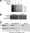
Ura3-Pcl5 is ubiquitinated by Hul5. (A) sub62 cells, either wild type or hul5Δ mutant, transformed with either KB1432 (Ura3-HA-Pcl5) or KB1432 (Ura3-HA) and either CUP1p-Myc-UBI or CUP1p-UBI (5) were induced for 4 h with 0.2 mM CuSO4. In a first stage, Ura3-Pcl5 was immunoprecipitated with an anti-HA antibody, and then the precipitate was solubilized in gel loading buffer and analyzed by SDS-PAGE/Western blotting for the presence of Myc-reactive proteins. (B) Strain KY1302 (sub62 hul5Δ) transformed with either KB1954 (HUL5), KB1955 (hul5 C878A), or vector (pRS315) and 5-fold dilutions of the resulting strains were spotted on synthetic complete medium without leucine and with or without uracil and incubated for 2 days (+uracil) or 3 days (−uracil). (C) Strain KY1302 was transformed with KB1169 (Ura3-Pcl5) and with either KB1954 (HUL5), KB1955 (hul5 C878A), or vector (pRS315). Ura3-Pcl5 degradation was followed by cycloheximide translation inhibition as for Fig. 1A.
FIG. 5.
FIG. 5.
Genetic interaction between hul5Δ and ubp6Δ in Ura3-Pcl5 processing. (A) Uracil prototrophy in the hul5Δ, ubp6Δ, and double mutants. Fivefold dilutions of the indicated mutants carrying plasmid KB1432 were spotted on synthetic complete medium with or without uracil and incubated for 2 days (+uracil) or 3 or 4 days (−uracil), as indicated, at 30°C. (B) The same strains were analyzed for Ura3-Pcl5 processing by cycloheximide chase as in Fig. 1A. (C) To quantitate the relative effect of Hul5 and Ubp6 on Ura3-Pcl5 processing, the same strains were subjected to [35S]methionine pulse-chase analysis as described in Materials and Methods, and the Ura3-Pcl5 full-length and processing products were detected with the anti-HA antibody. The arrows indicate the position of full-length Ura3-Pcl5, and the arrowheads indicate the position of the processed product. Quantitation of the protein bands is represented in the graphs. For the hul5Δ versus ubp6Δ hul5Δ experiment, the graph represents the average of results from four experiments; bars indicate standard deviations. Quantitation of the processing product was corrected for the smaller number of labeled residues as in Fig. 2A.
FIG. 6.
FIG. 6.
Partial processing of GFP-Pcl5 fusions. (A) Cycloheximide chase of GFP-Pcl5, GFP-Pcl5(71-229), and GFP-Pcl5(71-229) expressed from plasmids KB1943, KB1951, and KB1952 in wild-type (sub62) versus hul5Δ (KY1302) cells. The asterisk indicates a nonspecific band. C, no-GFP control. (B) [35S]methionine pulse-chase analysis of GFP-Pcl5 in wild-type versus hul5Δ cells. C, no-GFP control. (C) Quantitation of GFP-Pcl5 degradation versus processing (average of 2 experiments, included that shown in panel B). The percentage of the processing product is indicated relative to the full-length protein at t0 of the chase, corrected for the smaller number of methionines and cysteines (the residues labeled with 35S) in the processing product, namely, 9, versus 18 in the full-length GFP-Pcl5 fusion protein. Note that the processed band exceeds 100% in the hul5Δ mutant because the stable GFP band started accumulating during the pulse-labeling period.
FIG. 7.
FIG. 7.
Processive degradation of Gcn4(62-202)-Pcl5. Gcn4 T165A (62-202) was expressed from the CUP1 promoter of plasmid KB2012 in wild-type (sub62) cells versus hul5Δ (KY1302) cells. Degradation of the fusion protein was followed by [35S]methionine pulse-chase analysis. C, vector control.
FIG. 8.
FIG. 8.
Partial processing of Ura3-Pcl5 in the proteasomal ATPase mutant rpt2RF. (A) Cycloheximide-chase analysis of Ura3-Pcl5 processing in the hul5Δ, rpt2RF, and double mutants (strains KY1302, KY929, and KY1303, respectively). (B) Pulse-chase analysis of Ura3-Pcl5 using the same strains. (C) Pulse-chase analysis of Ura3-Pcl5 in KY929 and KY1302 versus Ura3-Pcl5(1-53) in wild-type cells. The two former strains were induced with 0.2 mM CuSO4, whereas the latter strain was induced with 10 μM CuSO4. Quantitation of the processing product in panels B and C was corrected for the smaller number of labeled residues as in Fig. 2A.

Similar articles

Cited by

References

    1. Aviram, S., E. Simon, T. Gildor, F. Glaser, and D. Kornitzer. 2008. Autophosphorylation-induced degradation of the Pho85 cyclin Pcl5 is essential for response to amino acid limitation. Mol. Cell. Biol. 28:6858-6869. - PMC - PubMed
    1. Chernova, T. A., K. D. Allen, L. M. Wesoloski, J. R. Shanks, Y. O. Chernoff, and K. D. Wilkinson. 2003. Pleiotropic effects of Ubp6 loss on drug sensitivities and yeast prion are due to depletion of the free ubiquitin pool. J. Biol. Chem. 278:52102-52115. - PubMed
    1. Crosas, B., J. Hanna, D. S. Kirkpatrick, D. P. Zhang, Y. Tone, N. A. Hathaway, C. Buecker, D. S. Leggett, M. Schmidt, R. W. King, S. P. Gygi, and D. Finley. 2006. Ubiquitin chains are remodeled at the proteasome by opposing ubiquitin ligase and deubiquitinating activities. Cell 127:1401-1413. - PubMed
    1. De Los Rios, P., A. Ben-Zvi, O. Slutsky, A. Azem, and P. Goloubinoff. 2006. Hsp70 chaperones accelerate protein translocation and the unfolding of stable protein aggregates by entropic pulling. Proc. Natl. Acad. Sci. U. S. A. 103:6166-6171. - PMC - PubMed
    1. Ellison, M. J., and M. Hochstrasser. 1991. Epitope-tagged ubiquitin. A new probe for analyzing ubiquitin function. J. Biol. Chem. 266:21150-21157. - PubMed

Publication types

MeSH terms

Substances