. 2010 Feb;30(4):922-34.
doi: 10.1128/MCB.01129-09. Epub 2009 Dec 14.

MAGUK-controlled ubiquitination of CARMA1 modulates lymphocyte NF-kappaB activity

Affiliations

MAGUK-controlled ubiquitination of CARMA1 modulates lymphocyte NF-kappaB activity

Miguel E Moreno-García et al. Mol Cell Biol. 2010 Feb.

Abstract

The adaptor protein CARMA1 is required for antigen receptor-triggered activation of IKK and JNK in lymphocytes. Once activated, the events that subsequently turn off the CARMA1 signalosome are unknown. In this study, we found that antigen receptor-activated CARMA1 underwent lysine 48 (K48) polyubiquitination and proteasome-dependent degradation. The MAGUK region of CARMA1 was an essential player in this event; the SH3 and GUK domains contained the main ubiquitin acceptor sites, and deletion of a Hook domain (an important structure for maintaining inactive MAGUK proteins) between SH3 and GUK was sufficient to induce constitutive ubiquitination of CARMA1. A similar deletion promoted the ubiquitination of PSD-95 and Dlgh1, suggesting that a conserved mechanism may control the turnover of other MAGUK family protein complexes. Functionally, we demonstrated that elimination of MAGUK ubiquitination sites in CARMA1 resulted in elevated basal and inducible NF-kappaB and JNK activation as a result of defective K48 ubiquitination and increased persistence of this ubiquitination-deficient CARMA1 protein in activated lymphocytes. The coordination of degradation with the full activation of the CARMA1 molecule likely provides an intrinsic feedback control mechanism to balance lymphocyte activation upon antigenic stimulation.


Figures

FIG. 1.
FIG. 1.
Degradation of stably expressed, constitutively active CARMA1 in human Ramos B cells. (A) Expression of myc epitope (CARMA1) and IKK-α/β in human Ramos B cells stably transduced with retroviral vectors expressing myc-CARMA1 or myc-CARMA1-ΔPRD and cis-linked IRES GFP. Expression of bicistronic GFP was analyzed by fluorescence-activated cell sorting (bottom). Solid histograms, untransduced cells; open histograms, myc-CARMA1-WT- or -ΔPRD-expressing cells. Numbers indicate the GFP median fluorescence intensity. (B) CARMA1-transduced Ramos cells were incubated with 50 μM CHX from 0 to 2 h, and WB analysis was carried out using antibodies for myc (CARMA1), IKKγ, and actin. (C) Densitometry of CARMA1 turnover in cells expressing myc-CARMA1-WT versus myc-CARMA1-ΔPRD (medians ± standard errors of the means of four independent experiments). Ratios of myc-CARMA1 versus actin levels quantified by fluorescence detection were used for normalization, and relative fold differences were determined using the zero time point set as 1. n.s., not significant; **, P ≤ 0.01; *, P ≤ 0.05 according to Student's t test.
FIG. 2.
FIG. 2.
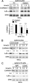
Degradation of endogenous CARMA1 in AR- or P/I-stimulated primary lymphocytes. Total splenocytes or splenic B cells were purified from adult C57/B6 mice, serum starved, and pretreated with 50 μM CHX. (A) Splenocytes were left unstimulated (DMSO) or stimulated with P/I for the indicated times. (Lower graph) Quantification of CARMA1 expression showing P/I-stimulated versus unstimulated cells. The graph shows means ± standard errors of the means from four independent experiments. (B) Splenocytes were left unstimulated (−) or stimulated with 10 μg/ml of both anti-CD3 and anti-CD28 for the indicated times. (C) Purified splenic B cells were stimulated with 10 μg/ml of anti-IgM or anti-CD40 (IC10) for the indicated times. In all experiments total CARMA1, IκB-α, and ERK expression levels were evaluated by immunoblotting (WB). Ratios of CARMA1 versus ERK levels were used for normalization, and fold differences were determined using the zero time point set as 1. n.s., no significant; **, P ≤ 0.01 according to Student's t test.
FIG. 3.
FIG. 3.
Constitutively active and endogenous CARMA1 are processed by the proteasome. (Α) Ramos B cells expressing myc-CARMA1-ΔPRD were either untreated (PBS or DMSO) or preincubated with lysosome inhibitors (NH4Cl or chloroquine) or proteasome inhibitors (MG132 or ALLN) for 20 min and subsequently treated with CHX or vehicle (DMSO) for 3 h. myc-CARMA1-ΔPRD, IκB-α, and actin were detected by WB. (B) Splenocytes were preincubated with DMSO or 100 μM proteasomal inhibitor ALLN or MG132 and then stimulated with P/I for the indicated times. Endogenous CARMA1, IκB-α, and ERK were detected by WB. Ratios of myc or endogenous CARMA1 versus actin or ERK levels were used for normalization, and relative fold differences were determined using the zero time point set as 1.
FIG. 4.
FIG. 4.
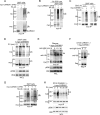
Constitutively active CARMA1-ΔPRD and activated CARMA1-WT are conjugated with K48-linked ubiquitin in 293T cells and B lymphocytes. (A) 293T cells were cotransfected with combinations of myc-CARMA1-ΔPRD and HA-Ub for 48 h. Cells were lysed with RIPA buffer (0.1% SDS, 0.5% Na-deoxycholate, 1% Triton X-100, and 250 mM NaCl) and myc-CARMA1-ΔPRD was immunoprecipitated with anti-myc antibodies and protein G beads (myc-IP). Ubiquitination was detected by anti-HA (Ub; brackets), and loading was detected by anti-myc WB. (B) 293T cells were transfected and processed as for panel A, with the exception that cells were lysed with RIPA containing 1% SDS and were diluted 10-fold to allow myc-CARMA1 immunoprecipitation (myc-IP) as described in Materials and Methods. (C) 293T cells were cotransfected for 48 h with combinations of myc-CARMA1 and HA-Ub and lysed with RIPA (0.1% SDS) or RIPA containing 1% SDS. myc-IP and WB were carried out as described for panel A. (D) 293T cells were cotransfected with myc-CARMA1 and HA-Ub. Cell were harvested and stimulated with P/I for the indicated times. Cells were processed and analyzed as for panel A. (E and F) Human Ramos B cells (25 × 106/condition) retrovirally transduced to stably express myc-CARMA1 (E) and CARMA1−/− DT40 B cells (4 × 10⁷/condition) with or without (-) Flag-CARMA1 expressed from the β-actin locus (F) were stimulated with anti-IgM for the indicated times, and then cells were lysed with RIPA. myc-IP or anti-Flag IP (flag-IP) was carried out, and CARMA1 ubiquitination in myc- and flag-IPs was detected using an anti-ubiquitin antibody (FK2). WB assays with anti-myc and anti-Flag antibodies were used as loading controls. (G) 293T cells were transfected with myc-CARMA1-ΔPRD and specific HA-Ub mutants (R48, Lys-48 mutated to Arg; K48, all Lys mutated to Arg except Lys-48; R63, Lys-63 mutated to Arg) in order to determine the type of ubiquitin chains formed. Cells were processed as for panel A. (H) CARMA1−/− DT40 cells retrovirally transduced to express myc-CARMA1 were stimulated with P/I for the indicated times, and endogenous K48-ubiquitination was detected using a specific anti-K48-linked ubiquitin antibody in myc-IPs. In all experiments the cells were preincubated for 1 h with 25 μM ALLN. (D, E, and H) Cell activation was analyzed by anti-pERK/ERK WB in WCL.
FIG. 5.
FIG. 5.
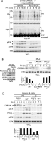
PKCβ controls CARMA1 ubiquitination and degradation in B cells. (A) CARMA1−/− DT40 cells retrovirally transduced to express myc-CARMA1 were preincubated with vehicle (DMSO) or 5 μM Ro-318425 (PKC inhibitor) for 30 min before being stimulated with P/I for the indicated times in the presence of 25 μM ALLN. Ubiquitination was observed in myc-IPs using anti-Ub, and loading was determined by using anti-myc antibody. The anti-myc WB was overexposed to show the anti-myc laddering. (B) CARMA1−/−, PKCβ−/−, or WT DT40 cells were left stimulated for the indicated times with P/I in the presence of CHX (right panel). (C) WT and PKCβ−/− splenic B cells were left unstimulated or stimulated with P/I for the indicated times in the presence of CHX. Cells were lysed with RIPA, and WCLs were tested for CARMA1, ERK, and PKCβ expression. For all panels, JNK and ERK phosphorylation levels were evaluated as specific activation markers. Asterisk, cross-reactivity of pJNK antibody with the pERK signal (A). Bar graphs represent CARMA1 expression relative to the loading control, ERK; results at the zero time points were set as 1.
FIG. 6.
FIG. 6.
The Hook domain in the MAGUK region suppresses CARMA1 ubiquitination at the GUK and SH3 domains. (A, upper panel) Diagram of the structural domains included in the 6×myc-tagged CARMA1 fragments (labeled A to G). 293T cells were cotransfected with HA-Ub and candidate 6×myc fragments. (B) Amino acid sequence alignment of the MAGUK regions from murine CARMA1, PSD-95, and Dlgh1. Amino acid identities are highlighted in black, and similarities are outlined in boxes. Bars beneath the sequences show the locations of the various domains. Blue, tripartite SH3 domain (the complete SH3 domain is formed spatially through protein folding rather than as a linear sequence); red, Hook segment; black, GUK domain. (C) 293T cells were cotransfected with HA-Ub and candidate 6×myc-tagged CARMA1 fragments. (D) 293T cells were cotransfected with HA-Ub and either myc-CARMA1-WT, -ΔPRD, or -ΔHook. (E). 293T cells were cotransfected with HA-Ub and either WT or ΔHook forms of myc-Dlgh1. (F) 293T cells were cotransfected with HA-Ub and either WT or ΔHook forms of myc-CARMA1 or myc-PSD-95. Cells were lysed with RIPA buffer (A to E) or with RIPA containing 1% SDS and then lysates were diluted 10-fold (F). myc-IPs were performed and ubiquitination and myc-tagged proteins were detected using anti-HA and anti-myc antibodies, respectively.
FIG. 7.
FIG. 7.
Elimination of ubiquitination of the MAGUK region reduces CARMA1 degradation and results in increased JNK and NF-κB activation. (A) 293T cells were cotransfected with HA-Ub and either myc-CARMA1-WT or a myc-CARMA1 mutant in which the lysine residues in the MAGUK region were replaced by arginines (myc-CARMA1-KR). Cells were harvested and stimulated with P/I for the indicated times. CARMA1 ubiquitination was detected as described previously using anti-HA and anti-K48 specific antibodies. Activation was detected using anti-pERK WB and WCL. (B) Retrovirally transduced CARMA1−/− DT40 cells were stimulated with P/I for the indicated times, and myc-CARMA1-WT (upper panels) or myc-CARMA1-KR (lower panels) degradation was analyzed by anti-myc WB in WCL. Cell activation and loading were assessed by anti-pERK/ERK WB. (C) WCLs from P/I-stimulated 293T cells transfected with WT- or KR-CARMA1 were generated and analyzed for JNK phosphorylation using polyclonal pJNK-specific antibodies. The bar graph represents the levels of pJNK relative to myc-CARMA1 expression. Zero time points from myc-CARMA1-transfected cells were set as 1. Data show averages ± standard errors of the means from three independent experiments. (D) Jurkat cells were cotransfected with reporter vector Igκ2-IFN-luciferase (NF-κB-Luc reporter), pRL-TK (transfection control), and DNA concentrations for myc-CARMA1-WT, myc-CARMA1-KR, or empty vector that resulted in similar expression of CARMA1 molecules. Cells were stimulated without or with CD3- and CD28-specific antibodies for 6 h. NF-κB-dependent luciferase activity normalized to the transfection control is shown (means ± standard errors of the means of three independent experiments). **, P ≤ 0.01; *, P ≤ 0.05 according to Student's t test. (Inset) myc and ERK WB for analysis of expression of myc-CARMA1-WT and myc-CARMA1-KR in 293T cells.
FIG. 8.
FIG. 8.
E3s target CARMA1 and downregulate CARMA1-dependent NF-κB activation but not its degradation. (A) 293T cells were cotransfected with myc-CARMA1-ΔPRD, HA-Ub, and candidate flag-tagged (f) E3s. HA (Ub) and myc (CARMA1) WB were assessed in myc-IPs. Expression of E3s was assessed in an anti-Flag WB assay in WCL. (B) 293T cells were cotransfected with myc-CARMA1-ΔPRD, f-cIAP2-WT, or the f-cIAP2-RING mutant. Ub and CARMA1 were detected in myc-IPs by anti-HA and anti-myc WB. WCL were analyzed to assess flag (cIAP2) expression level (bottom panel). (C) 293T cells were cotransfected with myc-CARMA1-WT and 2×Flag (f)-cIAP2, f-NEDD4, or f-Cbl-b and NF-κB and transfection control reporters. NF-κB-dependent luciferase activity was normalized to the transfection control. Data are means ± standard errors of the means of five independent experiments. For statistical analysis, Student's t test was used to compare cells expressing only CARMA1 versus those expressing CARMA1 plus specific E3s. **, P ≤ 0.05. Expression of CARMA1 and E3s was confirmed by WB of WCL using tag-specific antibodies. (D) Jurkat T cells were cotransfected for 48 h with NF-κB and control reporter genes and with various doses of myc-CARMA1-WT (266, 133, 66, and 33 ng) and f-cIAP2-wt, f-cIAP2-RING mutant, or empty vector. Unstimulated or anti-CD3ɛ/CD28-stimulated cells were assessed for NF-κB reporter activity. (E) Total splenocytes were pretreated with cIAP antagonist (BV6) for 1 h in order to promote cIAP1/2 turnover, and then stimulated with P/I for the indicated times in the presence of CHX and BV6. Endogenous CARMA1, cIAP1/2, and actin expression levels were analyzed by WB. CARMA1 fold differences were obtained from the CARMA1 versus actin ratios and normalized by setting the zero time points as 1.
FIG. 9.
FIG. 9.
Working model of coordinate CARMA1 degradation. CARMA1 exists predominantly in a double closed conformation. Upon cell activation, PKCβ/θ phosphorylate the PRD, promoting conformational changes and protein interactions that facilitate NF-κB and JNK activation. In parallel, conformational changes in the MAGUK region promote recruitment of one or various E3s to CARMA1, leading to K48-specific ubiquitination of the exposed GUK and SH3 domains followed by proteasome-dependent degradation of CARMA1. These combined events function coordinately to attenuate CARMA1-dependent NF-κB activation. The key E3s required for this process and the mechanisms that control Hook-dependent conformational changes are still unknown.

Similar articles

Cited by

References

    1. Bajpai, U. D., K. Zhang, M. Teutsch, R. Sen, and H. H. Wortis. 2000. Bruton's tyrosine kinase links the B cell receptor to nuclear factor κB activation. J. Exp. Med. 191:1735-1744. - PMC - PubMed
    1. Bertin, J., L. Wang, Y. Guo, M. D. Jacobson, J. L. Poyet, S. M. Srinivasula, S. Merriam, P. S. DiStefano, and E. S. Alnemri. 2001. CARD11 and CARD14 are novel caspase recruitment domain (CARD)/membrane-associated guanylate kinase (MAGUK) family members that interact with BCL10 and activate NF-kappa B. J. Biol. Chem. 276:11877-11882. - PubMed
    1. Egawa, T., B. Albrecht, B. Favier, M. J. Sunshine, K. Mirchandani, W. O'Brien, M. Thome, and D. R. Littman. 2003. Requirement for CARMA1 in antigen receptor-induced NF-kappa B activation and lymphocyte proliferation. Curr. Biol. 13:1252-1258. - PubMed
    1. Fields, E. R., B. J. Seufzer, E. M. Oltz, and S. Miyamoto. 2000. A switch in distinct I kappa B alpha degradation mechanisms mediates constitutive NF-kappa B activation in mature B cells. J. Immunol. 164:4762-4767. - PubMed
    1. Gaide, O., B. Favier, D. F. Legler, D. Bonnet, B. Brissoni, S. Valitutti, C. Bron, J. Tschopp, and M. Thome. 2002. CARMA1 is a critical lipid raft-associated regulator of TCR-induced NF-kappa B activation. Nat. Immunol. 3:836-843. - PubMed

Publication types

MeSH terms