. 2009 Dec 14;187(6):905-20.
doi: 10.1083/jcb.200908019.

Basal body stability and ciliogenesis requires the conserved component Poc1

Affiliations

Basal body stability and ciliogenesis requires the conserved component Poc1

Chad G Pearson et al. J Cell Biol. .

Abstract

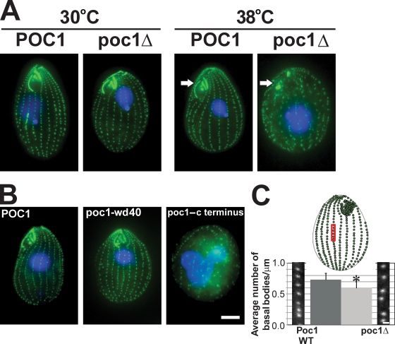
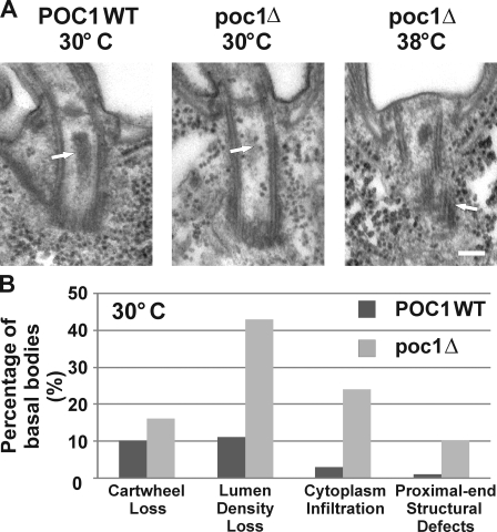
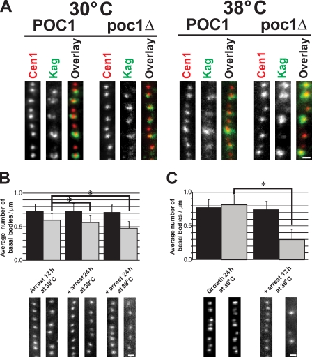
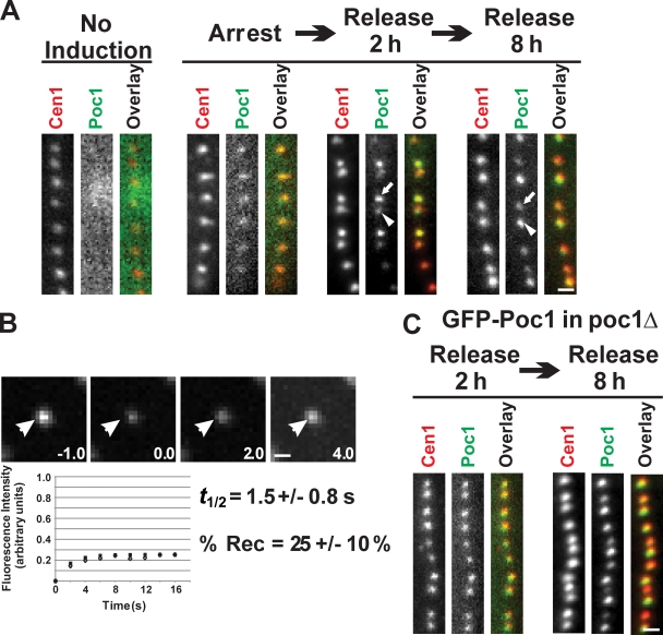
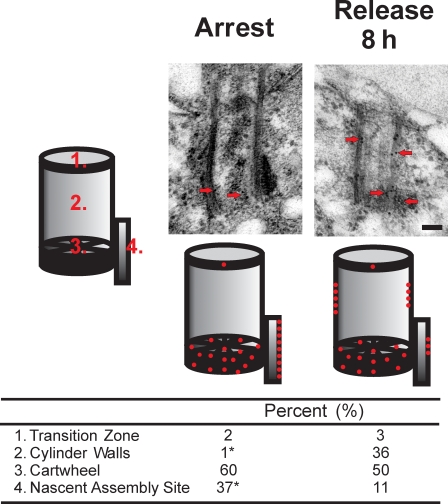
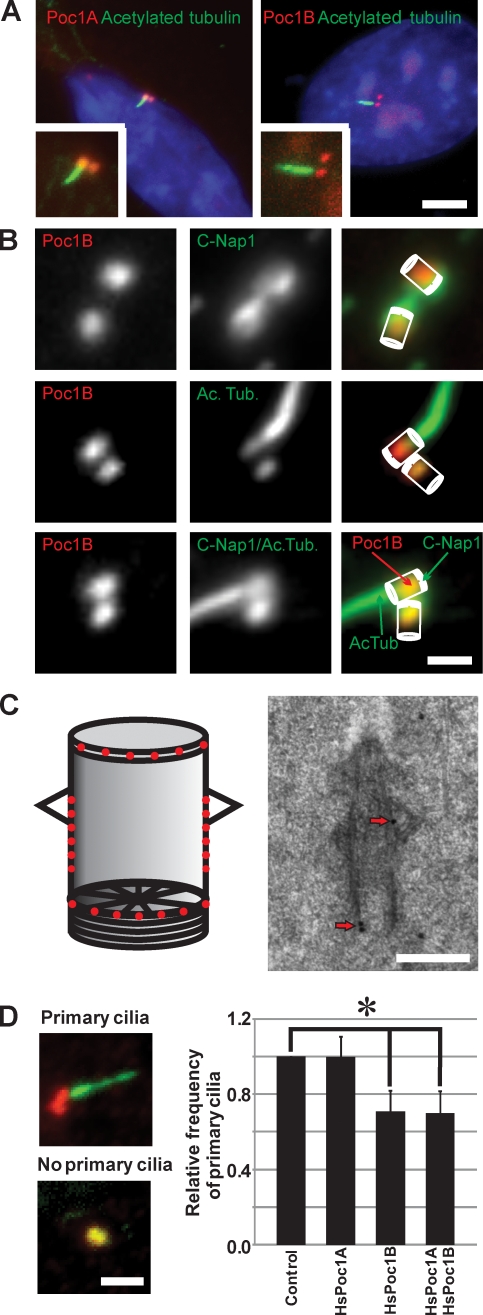
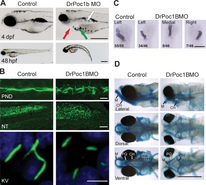
Centrioles are the foundation for centrosome and cilia formation. The biogenesis of centrioles is initiated by an assembly mechanism that first synthesizes the ninefold symmetrical cartwheel and subsequently leads to a stable cylindrical microtubule scaffold that is capable of withstanding microtubule-based forces generated by centrosomes and cilia. We report that the conserved WD40 repeat domain-containing cartwheel protein Poc1 is required for the structural maintenance of centrioles in Tetrahymena thermophila. Furthermore, human Poc1B is required for primary ciliogenesis, and in zebrafish, DrPoc1B knockdown causes ciliary defects and morphological phenotypes consistent with human ciliopathies. T. thermophila Poc1 exhibits a protein incorporation profile commonly associated with structural centriole components in which the majority of Poc1 is stably incorporated during new centriole assembly. A second dynamic population assembles throughout the cell cycle. Our experiments identify novel roles for Poc1 in centriole stability and ciliogenesis.


Figures

Figure 1.
Figure 1.
Basal body frequency and organization defects in poc1Δ. (A) Wild-type (POC1) and poc1 knockout (poc1Δ) T. thermophila cells were stained for basal bodies (green) and DNA (blue) after growing cells at either 30 or 38°C for 24 h. poc1Δ cells at 38°C exhibit a decreased frequency and disorganization of basal bodies and disruption of the oral apparatus (arrows). (B) poc1Δ cells with GFP-POC1 (aa 1–634), GFP-poc1-wd40 (aa 1–342), or GFP-poc1–c terminus (aa 343–634) at 38°C for 48 h. GFP-POC1 and the GFP-poc1-wd40 truncation rescues the lethality, oral apparatus defects, and basal body organization and frequency defects. (C) The frequency of basal bodies in poc1Δ cells is decreased in cells arrested by media starvation at 30°C for 12 h. The number of basal bodies in the medial 10 µm of cortical rows was measured to determine the mean number of basal bodies per micrometer. The red box is a representative region within the medial region of the T. thermophila cell selected for analysis. WT, wild type. *, P < 0.01; n > 150. Error bars indicate mean ± SD. Bars: (A and B) 10 µm; (C) 1 µm.
Figure 2.
Figure 2.
Poc1 loss causes basal body structural defects. (A) Basal body structural defects in poc1Δ cells at either 30 or 38°C for 24 h. Basal bodies in poc1Δ cells at 30°C exhibited a loss in the lumen electron density (arrows) and an infiltration of cytoplasm into the lumen (indicated by the ribosomal-like structures). (right) Defects in the triplet microtubules at the proximal end of basal bodies were observed at 30°C and were more frequently observed in the limited basal bodies at 38°C. Bar, 100 nm. (B) The relative frequency of the defects observed at 30°C. WT, wild type. n > 100 basal bodies.
Figure 3.
Figure 3.
Poc1 is required for basal body stability. (A) New basal bodies assemble in the absence of Poc1. All basal bodies (centrin, red) were observed relative to mature basal bodies (K antigen, green). Newly formed basal bodies (no K antigen) were found at all conditions. (B) Basal bodies are less stable in poc1Δ cells. Cells were arrested at 30°C for 12 h (left) to create a uniform cell cycle population, and the arrest was maintained either at 30 (middle) or 38°C (right) for an additional 24 h. Basal body frequency was quantified for each condition by counting the number of centrin-stained basal bodies per micrometer. (C) Basal bodies assembled in poc1Δ cells at 38°C are less stable than at 30°C. Cells were grown at 38°C for 24 h before arrest for 12 h at 38°C. Black bars, wild-type POC1; gray bars, poc1Δ. *, P < 0.01; n > 150. Error bars indicate mean ± SD. Bars, 1 µm.
Figure 4.
Figure 4.
Poc1 exhibits both dynamic and stable incorporation at basal bodies. (A) GFP-Poc1 protein incorporation during basal body assembly. Noninduced cells exhibited no fluorescence signal. GFP-Poc1 expressed for 2 h in cells arrested for 12 h to inhibit basal body assembly. Low fluorescence uniformly incorporated at all basal bodies. Cells were released from the arrest for 2 h in the presence of GFP-Poc1. Dim GFP-Poc1 signal was found at existing, mature basal bodies (arrowhead), whereas newly assembled basal bodies contained bright GFP-Poc1 (arrow). Uniform anticentrin staining was observed at all basal bodies. 8 h after release, the majority of the basal bodies were brightly labeled. In contrast to the 2-h release, at 8 h, new basal bodies in dividing cells were dim (arrow) compared with old basal bodies (arrowhead). (B) FRAP to quantify the dynamic fraction of GFP-Poc1 in poc1Δ cells. After photobleaching, fluorescence recovery was followed. (bottom) Open diamonds indicate the mean fluorescence recovery relative to the best-fit single exponential recovery (filled squares). Arrowheads indicate the photobleached basal body. (C) GFP-Poc1 decorates all basal bodies uniformly upon expression in cycling cells for 2 h in a poc1Δ strain. Bars, 1 µm.
Figure 5.
Figure 5.
Dynamic and stable populations of Poc1 incorporation. IEM of GFP-Poc1 was visualized in cells arrested by starvation (arrest) and normally cycling cells (release 8 h). In arrested cells, GFP-Poc1 incorporates at only the cartwheel and the nascent site of assembly. In contrast, GFP-Poc1 assembles at basal body cylinder walls in addition to the cartwheel and nascent site when expressed in cycling cells with new basal body assembly in the presence of GFP-Poc1. The relative frequency of gold label at the nascent site of basal body assembly decreases in cycling cells compared with arrested cells. Red dots indicate the relative distribution of gold particles (n > 150). *, P < 0.01 by χ2 test. Bar, 100 nm.
Figure 6.
Figure 6.
Human Poc1B is required for ciliogenesis. (A) Basal body localization of mCherry-Poc1A or mCherry-Poc1B (red) was visualized relative to ciliary axonemes labeled with anti-acetylated tubulin (green) in ciliated human RPE-1 cells. Insets indicate a magnified view. Bar, 5 µm. (B) HsPoc1B localized predominantly to the proximal end of basal bodies. Poc1B (red; CO200) was colocalized relative to C-Nap1 (green; basal body proximal end marker) and acetylated tubulin (ac tub; green; basal body and cilia marker). Bar, 1 µm. (C) IEM localized HsPoc1B to the transition zone (23%), cylinder walls (49%), and cartwheel (28%). (left) Schematic showing the relative distribution of gold particles. (right) A representative localization image is shown. Arrows indicate immunogold label. Bar, 200 nm. (D) Depletion of HsPoc1B inhibits primary cilia formation. Cilia axonemes were labeled with anti-acetylated tubulin (green), and basal bodies were labeled with γ-tubulin (red). The presence or absence of primary cilia was scored to determine the relative frequency of primary cilia relative to control siRNA. Bar, 2 µm. P < 0.01; n > 800 cells; n = 4. Error bars indicate mean ± SD.
Figure 7.
Figure 7.
Zebrafish Dr.poc1B morphants exhibit ciliopathy defects. (A) Dr.poc1B morphants (DrPoc1B MO) display smaller eyes, heart edemas (red arrow), mislocalized melanocytes (green arrow), truncated bodies, and kidney cysts (white arrow) at 4 d post fertilization (dpf). Bars, 0.5 mm. (B) Cilia (green) of Dr.poc1B morphant embryos at 48 hpf are disorganized and appear shorter within the pronephretic duct (PND) and are expanded within the posterior neural tube (NT). Short cilia were visible in the KV at 12 hpf (Dr.poc1BATG MO, 3.6 ± 0.9 µm; Dr.poc1BSPL MO, 3.6 ± 0.9 µm; control, 6.2 ± 0.8 µm; n > 100; P < 0.001). Bars: (top) 10 µm; (middle) 25 µm; (bottom) 5 µm. (C) Heart laterality defects were observed in Dr.poc1B morphants at 48 hpf. Using in situ hybridization to cmlc2, heart loops were visualized in flat-mounted sections. The ratios indicate the relative frequency of heart positioning. Bar, 0.5 mm. (D) Fish were stained with Alcian blue to visualize cartilage and bone. Morphants displayed craniofacial abnormalities consistent with the ciliopathy phenotype. Dr.poc1B morphants exhibit pharyngeal arch and jaw defects at 6 d post fertilization. PQ, palatoquadrate; M, Meckel's cartilage; CH, ceratohyal; VPA, visceral pharyngeal arches. Bar, 0.5 mm.

Similar articles

Cited by

References

    1. Abal M., Keryer G., Bornens M.. 2005. Centrioles resist forces applied on centrosomes during G2/M transition. Biol. Cell. 97:425–434. 10.1042/BC20040112 - DOI - PubMed
    1. Andersen J.S., Wilkinson C.J., Mayor T., Mortensen P., Nigg E.A., Mann M.. 2003. Proteomic characterization of the human centrosome by protein correlation profiling. Nature. 426:570–574. 10.1038/nature02166 - DOI - PubMed
    1. Avidor-Reiss T., Maer A.M., Koundakjian E., Polyanovsky A., Keil T., Subramaniam S., Zuker C.S.. 2004. Decoding cilia function: defining specialized genes required for compartmentalized cilia biogenesis. Cell. 117:527–539. 10.1016/S0092-8674(04)00412-X - DOI - PubMed
    1. Azimzadeh J., Bornens M.. 2004. The centrosome in evolution. In Centrosomes in Development and Disease. Nigg E.A., editor. Wiley-VCH, Weinheim, Germany. 431 pp.
    1. Badano J.L., Mitsuma N., Beales P.L., Katsanis N.. 2006. The ciliopathies: an emerging class of human genetic disorders. Annu. Rev. Genomics Hum. Genet. 7:125–148. 10.1146/annurev.genom.7.080505.115610 - DOI - PubMed

Publication types

MeSH terms