. 2010 Mar;184(3):717-29.
doi: 10.1534/genetics.109.111344. Epub 2009 Dec 14.

Drosophila Raf's N terminus contains a novel conserved region and can contribute to torso RTK signaling

Affiliations

Drosophila Raf's N terminus contains a novel conserved region and can contribute to torso RTK signaling

Jian Ding et al. Genetics. 2010 Mar.

Abstract

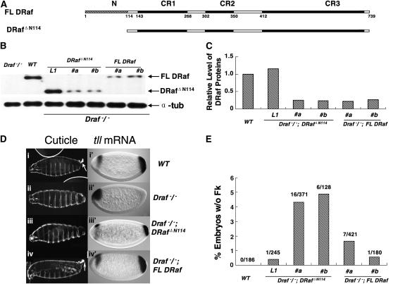
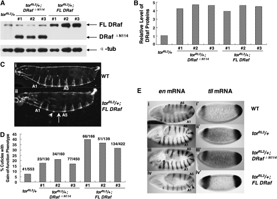
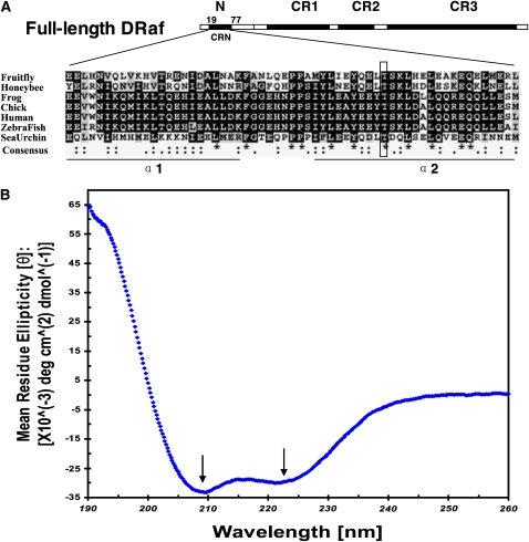
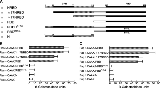
Drosophila Raf (DRaf) contains an extended N terminus, in addition to three conserved regions (CR1-CR3); however, the function(s) of this N-terminal segment remains elusive. In this article, a novel region within Draf's N terminus that is conserved in BRaf proteins of vertebrates was identified and termed conserved region N-terminal (CRN). We show that the N-terminal segment can play a positive role(s) in the Torso receptor tyrosine kinase pathway in vivo, and its contribution to signaling appears to be dependent on the activity of Torso receptor, suggesting this N-terminal segment can function in signal transmission. Circular dichroism analysis indicates that DRaf's N terminus (amino acids 1-117) including CRN (amino acids 19-77) is folded in vitro and has a high content of helical secondary structure as predicted by proteomics tools. In yeast two-hybrid assays, stronger interactions between DRaf's Ras binding domain (RBD) and the small GTPase Ras1, as well as Rap1, were observed when CRN and RBD sequences were linked. Together, our studies suggest that DRaf's extended N terminus may assist in its association with the upstream activators (Ras1 and Rap1) through a CRN-mediated mechanism(s) in vivo.


Figures

F<sc>igure</sc> 1.—
Figure 1.—
Rescue of posterior structures in embryos derived from Draf/ female germ cells by expression of full-length DRaf or truncated DRaf ΔN114 transgenes. (A) Schematic representations of full-length DRaf (FL DRaf) with 739 amino acids and truncated DRafΔN114 proteins. In addition to the three conserved regions (CR1, CR2, and CR3), FL DRaf has an extended N terminus. (B) Western analysis of embryonic DRaf proteins from eggs produced by Draf 11-29/Draf 11-29 (Draf/), wild type (WT), Draf/; DRaf ΔN114 (three independent lines, L1 with ∼1× endogenous DRaf level, #a and #b with ∼1/4 endogenous DRaf level) and Draf/; FL DRaf (two independent lines, #a and #b with ∼1/4 endogenous DRaf level) germline clone bearing females. Lysate was prepared from eggs at 0–3 hr after egg deposition. Full-length DRaf (∼90 kDa) and DRafΔN114 (∼77 kDa) proteins are denoted by arrows. Lysate of eggs from Draf/ germ cells was used as a negative control. α-Tubulin (α-tub) was used as the loading control. (C) A bar graph representing relative levels of DRaf proteins normalized with α-tubulin. (D) Cuticles of mature embryos derived from wild type (WT), Draf/, Draf/; DRaf ΔN114, and Draf/; FL DRaf female germ cells (left). Accumulation of tll mRNA in cellular blastoderm embryos was detected by in situ hybridization (right). Posterior tll expression is solely dependent on the Torso pathway and used as a marker for Torso RTK signaling. Anterior expression of tll is regulated by another pathway(s) in addition to Torso signaling, is more complex, and is used as an internal control for staining here. Wild-type embryos show (i) normal Filzkörper (Fk) structure (arrow), (i′) tll mRNA accumulation at the posterior (∼0–15% embryo length, EL), and an anterior head “stripe” (∼75–85% EL). Embryos derived from Draf/ germ cells lack (ii) posterior structures (A8 denticle belt, Filzkörper) and (ii′) posterior tll mRNA expression. (iii) An embryonic cuticle derived from Draf/; DRaf ΔN114 germ cell lacks the Filzkörper structure. (iii′) A reduced posterior tll expression domain is at ∼0–8% EL in an embryo from Draf/; DRaf ΔN114 germline clone bearing mother. (iv) Filzkörper structure (arrow) and (iv′) normal expression pattern of tll mRNAs are rescued by FL DRaf expression for embryos derived from Draf/; FL DRaf maternal germ cells. (E) A bar graph showing the percentage of embryos without Fk structures. When expressed at low maternal level (∼1/4 endogenous level), embryos without Fk were found more often for those that inherited truncated DrafΔN114 rather than full-length DRaf proteins (χ2 = 9.91976318, P < 0.01).
F<sc>igure</sc> 2.—
Figure 2.—
Verification of DRaf protein quantitation assays and stability of DRaf proteins in early embryos. (A) Three samples representing lystes of 6, 12, and 18 eggs for each line (Draf/; DRaf ΔN114#a and Draf/; FL DRaf #a) were loaded for Western blot analysis. Full-length DRaf (∼90 kDa) and DRafΔN114 (∼77 kDa) proteins are denoted by arrows. Lysate of eggs from Draf 11-29/Draf 11-29 (Draf/) germ cells was used as a negative control. α-Tubulin levels were probed as a loading control. (B) Bar graph showing relative intensity of DRaf (solid bar) and α-tubulin (shaded bar) bands. (C) A bar graph depicting normalized DRaf protein level from A. (D) Western analysis of embryonic DRaf proteins from eggs collected at 0–1, 1–2, and 2–3 hr after deposition and produced by Draf/, Draf/; DRafΔN114 (line #a) and Draf/; FL DRaf (line #a) germline-bearing females. Full-length DRaf (∼90 kDa) and DRafΔN114 (∼77 kDa) proteins are denoted by arrows. α-Tubulin was used as the loading control. (E) Normalized DRaf protein level from D is shown in this bar graph depicting the stable accumulation of these DRaf proteins.
F<sc>igure</sc> 3.—
Figure 3.—
Gain-of-function effects of torRL3 are differentially enhanced by expression of FL DRaf and DRaf ΔN114 transgenes. (A) Western analysis of embryonic DRaf proteins from eggs (0–3 hr) produced by torRL3/+, torRL3/+; DRaf ΔN114 (three independent lines, #1, #2, and #3), or torRL3/+;FL DRaf (three independent lines, #1, #2, and #3) females at 29°. Full-length DRaf (∼90 kDa) and DRafΔN114 (∼77 kDa) proteins are denoted by arrows. α-Tubulin was used as the loading control. (B) Normalized DRaf protein level from A is shown as a bar graph. (C) Cuticles of mature embryos are shown. (i) A wild-type (WT) embryo exhibits normal cuticle pattern with 8 abdominal denticle belts. (ii) An embryonic cuticle derived from torRL3/+; FL DRaf mother has one broken abdominal denticle band (arrow head) and is missing one central abdominal denticle belt (arrow). (D) Percentage of embryonic cuticles with gain-of-function phenotypes is shown. Gain-of-function effects of torRL3 were differentially enhanced by FL DRaf and DRafΔN114 proteins (χ2 = 51.063837, P < 0.001). (E) Expression of engrailed (en) at approximately stage 11 (left) and accumulation of tailless (tll) mRNA at cellular blastoderm stage (right) in embryos from WT, torRL3/+, torRL3/+;DRaf ΔN114, or torRL3/+;FL DRaf mothers: Examples of embryos derived from (i) WT, (ii) torRL3/+, and (iii) torRL3/+;DRaf ΔN114 mothers exhibit normal en mRNA pattern with three thoracic (T1–T3) and nine abdominal (A1–A9) expression stripes. (iv) An embryo from a torRL3/+; FL DRaf mother is shown with partial deletion of en stripes (arrow) in a region that gives rise to central abdominal segmental pattern. Examples of embryos derived from (i′) WT and (ii) torRL3/+ mothers exhibiting a normal tll mRNA pattern. (iii′) An embryo from a torRL3/+; DRaf ΔN114 mother shows slightly expanded posterior expression domain of tll. (iv′) An embryo derived from torRL3/+;FL DRaf females exhibits expanded domain of tll expression for both anterior and posterior regions.
F<sc>igure</sc> 4.—
Figure 4.—
Effects of FL DRaf and DRafΔN114 expression on posterior development in embryos derived from trk 1/trk 1 mothers. (A) Western analysis of embryonic DRaf proteins from eggs (0–3 hr) produced by Draf 11-29/Draf 11-29 (Draf/), wild type (WT), trk 1/trk 1 (trk/), trk/; DRaf ΔN114/DRaf ΔN114 (three lines, #1/#3, #2/#2, and #3/#3), and trk/; FL DRaf/FL DRaf (three lines, #1/#1, #2/#2, and #3/#3) females. Full-length DRaf (∼90 kDa) and DRafΔN114 (∼77 kDa) proteins are denoted by arrows. Embryonic lysate from Draf/ germline clone females was used as a negative control. α-Tubulin was used as the loading control. (B) Normalized DRaf protein level from A is shown in the bar graph. (C) Representative cuticles of mature embryos derived from wild type (WT), trk/, trk/; DRaf ΔN114/DRaf ΔN114, or trk/; FL DRaf/FL DRaf females are shown (left). Accumulation of tll (right) mRNAs was detected by in situ hybridization. (i) A wild-type (WT) embryo has normal cuticle pattern with eight abdominal denticle belts and Filzkörper structure. (i′) A WT embryo at cellular blastoderm stage exhibits a normal posterior expression domain of tll. (ii) Cuticle of a mature embryo from a trk/ mother is missing posterior structures (A8 segment, Filzkörper). (ii′) A cellular blastoderm embryo from a trk/ mother lacks posterior tll expression. (iii) An embryonic cuticle from a trk/; DRaf ΔN114/DRaf ΔN114 mother lacks posterior structures (A8 denticle belt, Filzkörper). (iii′) A cellular blastoderm embryo from a trk/; DRaf ΔN114/DRaf ΔN114 mother lacks posterior expression of tll mRNA. Expression of FL DRaf (iv) restores the A8 denticle belt (arrow) and (iv′) posterior tll expression (arrow) in embryos lacking maternal Trk activity. (D) Effect of FL DRaf or DRaf ΔN114 transgene expression on A8 denticle development in embryos derived from trk/ mothers (percentage of embryonic cuticles with A8 denticle belt). Shown are results using transgenic DRaf ΔN114 or FL DRaf lines that express DRaf protein at similar levels. Expression of exogenous FL DRaf, but not DRafΔN114, results in partial rescue of A8 denticle belt in some embryos derived from trk/ mothers (χ2 = 82.8574882, P < 0.001).
F<sc>igure</sc> 5.—
Figure 5.—
The N terminus of DRaf contains a novel conserved region and has a high content of helical secondary structure. (A) Drosophila Raf (NP_525047; 739 amino acids) has in addition to its three conserved regions (CR1–CR3), an extensive N terminus. A novel region (amino acids 19–77) within the N terminus is conserved in honeybee Raf (Apis mellifera XP_396892), frog BRaf (Xenopus laevis AAU29410), chicken C-Rmil (Gallus gallus CAA47436), human BRaf (Homo sapiens NP_004324), zebrafish BRaf (Danio rerio BAD16728), and sea urchin BRaf (Strongylocentrotus purpuratus XP_781094) and termed conserved region N-terminal (CRN). Sequences of CRN were aligned using ClustalW (identities were denoted as *; strong and weak similarities were denoted as : and .," respectively, in consensus line, http://www.ebi.ac.uk/tools/clustalw/), and the conserved residues were shaded using BOXSHADE (identities, solid; similarities, shaded, http://www.ch.embnet.org/software/BOX_form.html). Secondary structure prediction with GORV indicates CRN has the propensity to form two α-helices (α1 and α2). The putative PKC phosphorylation site DRaf's Thr60 is framed. (B) Circular dichroism (CD) spectral measurement of DRaf's N terminus (amino acids 1–117) in vitro: The bilobed spectrum (arrows, local minima at ∼209.4 nm and at ∼221.4 nm) indicative of helical secondary structure is shown.
F<sc>igure</sc> 6.—
Figure 6.—
Effects of the extended N terminus of DRaf on Ras1 and Rap1 binding. (A) Schematic representations of different DRaf constructs used for yeast two-hybrid analysis. (B) Interactions between DRaf's RBDs and Ras1ΔCAAX: Removal of CRN or the entire N-terminal region reduces Ras1ΔCAAX binding (P < 0.05, t-test). (C) Interactions between DRaf's RBDs and Rap1ΔCAAX: Removal of CRN or the entire N-terminal region reduces Rap1ΔCAAX binding (P < 0.05, t-test).

Similar articles

Cited by

References

    1. Baek, K.-H., J. R. Fabian, F. Sprenger, D. K. Morrison and L. Ambrosio, 1996. The activity of D-raf in torso signal transduction is altered by serine substitution, N terminal deletion and membrane targeting. Dev. Biol. 175 191–204. - PubMed
    1. Blom, N., S. Gammeltoft and S. Brunak, 1999. Sequence- and structure-based prediction of eukaryotic protein phosphorylation sites. J. Mol. Biol. 294 1351–1362. - PubMed
    1. Brennan, C. A., and K. Moses, 2000. Determination of Drosophila photoreceptors: timing is everything. Cell. Mol. Life Sci. 57 195–214. - PMC - PubMed
    1. Casanova, J., M. Furriols, C. A. Mccormick and G. Struhl, 1995. Similarities between trunk and spätzle, putative extracellular ligands specifying body pattern in Drosophila. Genes Dev. 9 2539–2544. - PubMed
    1. Chong, H., L. Jeeyong and K. L. Guan, 2001. Positive and negative regulation of Raf kinase activity and function by phosphorylation. EMBO J. 20 3716–3727. - PMC - PubMed

MeSH terms