. 2010 Mar;184(3):613-28.
doi: 10.1534/genetics.109.110338. Epub 2009 Dec 14.

A single unpaired and transcriptionally silenced X chromosome locally precludes checkpoint signaling in the Caenorhabditis elegans germ line

Affiliations

A single unpaired and transcriptionally silenced X chromosome locally precludes checkpoint signaling in the Caenorhabditis elegans germ line

Aimee Jaramillo-Lambert et al. Genetics. 2010 Mar.

Abstract

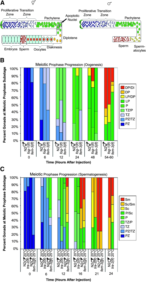
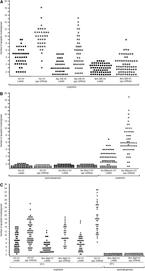
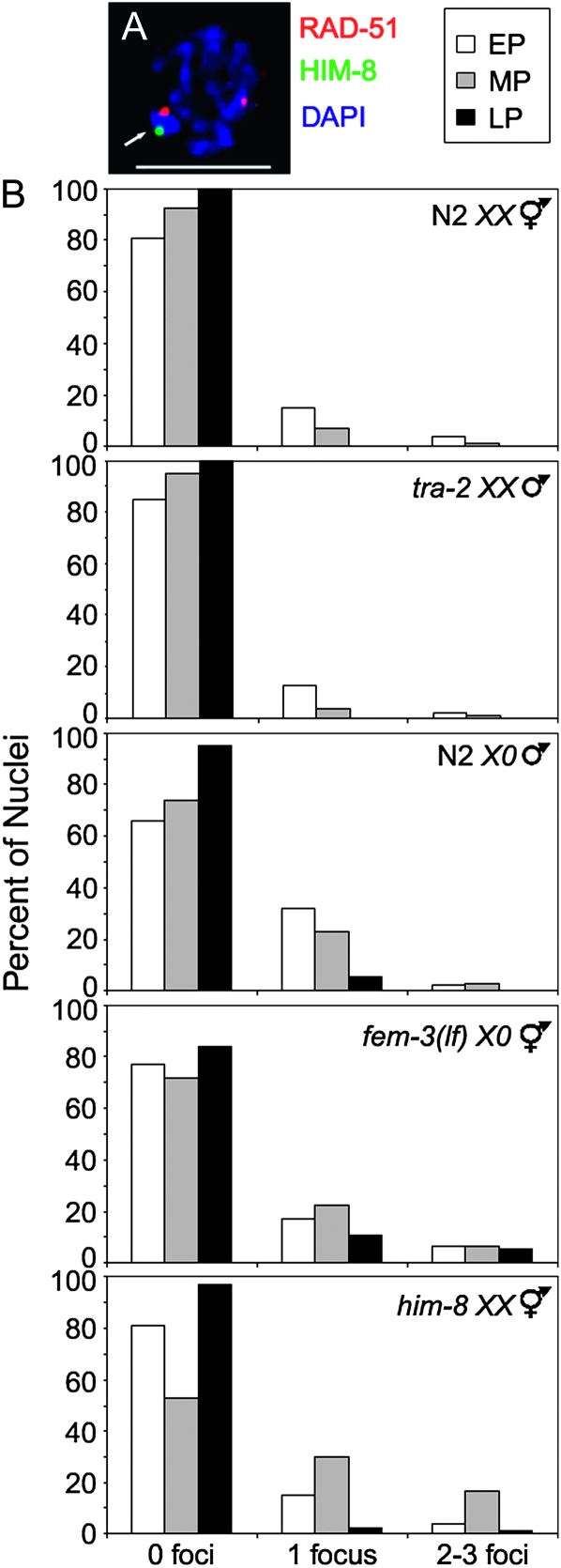
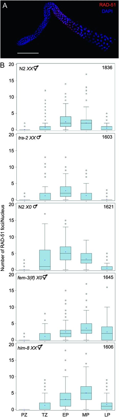
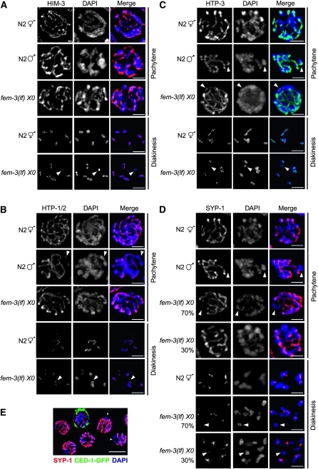
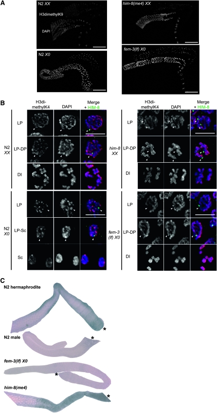
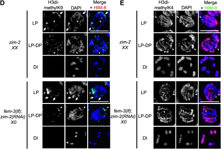
In many organisms, female and male meiosis display extensive sexual dimorphism in the temporal meiotic program, the number and location of recombination events, sex chromosome segregation, and checkpoint function. We show here that both meiotic prophase timing and germ-line apoptosis, one output of checkpoint signaling, are dictated by the sex of the germ line (oogenesis vs. spermatogenesis) in Caenorhabditis elegans. During oogenesis in feminized animals (fem-3), a single pair of asynapsed autosomes elicits a checkpoint response, yet an unpaired X chromosome fails to induce checkpoint activation. The single X in males and fem-3 worms is a substrate for the meiotic recombination machinery and repair of the resulting double strand breaks appears to be delayed compared with worms carrying paired X chromosomes. Synaptonemal complex axial HORMA domain proteins, implicated in repair of meiotic double strand breaks (DSBs) and checkpoint function, are assembled and disassembled on the single X similarly to paired chromosomes, but the central region component, SYP-1, is not loaded on the X chromosome in males. In fem-3 worms some X chromosomes achieve nonhomologous self-synapsis; however, germ cells with SYP-1-positive X chromosomes are not preferentially protected from apoptosis. Analyses of chromatin and X-linked gene expression indicate that a single X, unlike asynapsed X chromosomes or autosomes, maintains repressive chromatin marks and remains transcriptionally silenced and suggests that this state locally precludes checkpoint signaling.


Figures

F<sc>igure</sc> 1.—
Figure 1.—
Meiotic prophase progression is dependent on germ-line sex. (A) Cartoon of hermaphrodite (left) and male (right) gonad arms. Distal end is capped by somatic distal tip cell(s) (green) and contains a population of proliferating germ cells (dark blue). As cells move proximally they enter meiotic prophase (transition zone-leptotene/zygotene; light blue crescents) and then progress through pachytene (green), diplotene (yellow), and diakinesis (red). Initially, hermaphrodites form sperm (small red circles) and then switch to oocyte production (light blue with red centers) beginning at L4 stage. After switch to oocyte production, cells in the pachytene region of the germ line undergo physiological apoptosis (circled green nuclei). In males, germ-line cells differentiate into spermactocytes (yellow triangles) and then sperm (red circles); no apoptosis occurs. (B) Meiotic prophase timing in sex determination mutants undergoing oogenesis. Percentage of gonads displaying labeled nuclei at indicated stages is shown. Proliferative zone (PZ), transition zone (TZ), pachytene (P), late pachytene (LP), diplotene (DP), and diakinesis (DI). Number of gonads examined at each time point were N2 XX: 0 hr = 9, 6 hr = 14, 12 hr = 10, 24 hr = 17, 48 hr = 9, and 54–60 hr = 9; fog-1(q325) X0: 0 hr = 7, 6 hr = 9, 12 hr = 9, 24 hr = 9, 48 hr = 11, and 54–60 hr = 8; and fem-3(e1996) X0: 0 hr = 10, 6 hr = 12, 12 hr = 8, 24 hr = 12, 48 hr = 10, and 54–60 hr = 18. (C) Meiotic prophase timing in sex determination mutants undergoing spermatogenesis. Percentage of gonads with labeled nuclei at indicated stages is shown. Proliferative zone (PZ), transition zone (TZ), pachytene (P), spermatocytes (Sc), and sperm (Sm). Number of gonads examined at each time point were N2 X0 20°: 0 hr = 16, 6 hr = 8, 12 hr = 16, 16 hr = 19, 21 hr = 10, and 24 hr = 9; tra-2(e1095) XX 20°: 0 hr = 8, 6 hr = 8, 12 hr = 9, 16 hr = 8, 21 hr = 9, and 24 hr = 8; N2 X0 25°: 0 hr = 7, 6 hr = 7, 12 hr = 13, 16 hr = 7, and 21 hr = 4 (15/19 gonads no label); and fem-3(q20) XX 25°: 0 hr = 10, 6 hr = 9, 12 hr = 8, 16 hr = 10, and 21 hr = 8.
F<sc>igure</sc> 2.—
Figure 2.—
Physiological and checkpoint-activated apoptosis are dependent on germ-line sex. (A) Scatterplot depicting number of apoptotic bodies detected in germ lines of N2 XX, fog-1(q325) X0, and fem-3(e1996) X0 animals (by AO staining and DIC). Apoptosis was scored in adult animals 24 hr post-L4. Y-axis value for each point represents number of apoptotic bodies/gonad. Total number of gonads examined for each: N2 XX L4440 N = 50, N2 XX syp-1(RNAi) N = 52, fog-1(q325) X0 L4440 N = 48, fog-1(q325) X0 syp-1(RNAi) N = 50, fem-3(e1996) X0 L4440 N = 60, and fem-3(e1996) X0 syp-1(RNAi) N = 66. (B) Scatterplot depicting number of apoptotic bodies detected in germ lines of N2 X0, tra-2 XX, or tra-2/dpyunc XX worms (by AO staining and DIC). Apoptosis was scored in adult animals 48 hr post-L4. Y-axis value for each point represents number of apoptotic bodies/gonad. Total number of gonads examined for each: N2 X0 L4440 N = 37, N2 X0 syp-1(RNAi) N = 28, tra-2(e1095) XX L4440 N = 44, tra-2(e1095) XX syp-1(RNAi) N = 44, tra-2(e1095)/dpyunc XX L4440 N = 70, and tra-2(e1095)/dpyunc XX syp-1(RNAi) N = 57. (C) Scatterplot depicting number of apoptotic bodies detected in gonads of N2 XX and fem-3(q22) XX worms by CED-1∷GFP fluorescence, for both control (L4440) and syp-1(RNAi). fem-3(q22) is temperature sensitive and each experiment was performed at both 15° and 25°. Apoptotic bodies were scored in adult animals 48 hr post-L3. Y-axis values represent number of apoptotic bodies/gonad. Total number of gonads examined for 15°: N2 XX L4440 N = 50, N2 XX syp-1(RNAi) N = 65, fem-3(q22) XX L4440 N = 48, and fem-3(q22) XX syp-1(RNAi) N = 31. Total number of gonads examined for 25°: N2 XX L4440 N = 44, fem-3(q22) X0 L4440 N = 36, N2 XX syp-1(RNAi) N = 38, and fem-3(q22) XX syp-1(RNAi) N = 37.
F<sc>igure</sc> 3.—
Figure 3.—
Abundance of RAD-51 foci on X chromosome(s). (A) Nucleus showing RAD-51 focus on X (arrow). Nucleus labeled with α-RAD-51 (red) and α-HIM-8 (green), counterstained with DAPI (blue). Scale bar, 5 μm. (B) Histograms representing quantification of RAD-51 foci on X chromosome(s) in N2 XX, tra-2 XX males, N2 X0, fem-3(e1996) X0, and him-8(me4) XX. Y-axis indicates percentage of nuclei that contain 0, 1, or 2–3 RAD-51 foci per X chromosome for early pachytene (white), midpachytene (gray), and late pachytene (black).
F<sc>igure</sc> 4.—
Figure 4.—
Assembly and removal of RAD-51 foci during meiotic prophase progression. (A) Image of adult N2 X0 gonad stained with DAPI (blue) and α-RAD-51 (red). Scale bar, 50 μm. (B) Quantification of RAD-51 focus formation in N2 XX, tra-2(e1095) XX, N2 X0, fem-3(e1996) X0, and him-8(me4) XX. Gonads were divided into prophase substages and nuclei assigned to each region on the basis of morphology and location. Graphs display box-whisker plots of focus numbers. X-axis indicates meiotic prophase stages: Proliferative zone (PZ), transition zone (TZ), early pachytene (EP), midpachytene (MP), and late pachytene (LP); y-axis indicates number of RAD-51 foci/nucleus. Center horizontal line of each box indicates the median measurements; lines extending above and below boxes indicate standard deviation and outliers indicate the entire range of measurements. Numbers of nuclei observed for each strain are indicated in upper right.
F<sc>igure</sc> 5.—
Figure 5.—
Loading of SC axial and central elements onto the X chromosome(s). Immunolocalization of (A) HIM-3 (red), (B) HTP-1/2 (red), (C) HTP-3 (green), and (D) SYP-1 (red) counterstained with DAPI (blue) in nuclei from N2 XX, N2 X0, and fem-3(e1996) X0 germ lines. Arrowheads point to X chromosome determined by lack of localization of SYP-1 or by size of DAPI staining body. Scale bar, 2 μm. (E) SYP-1 (red) and CED-1-GFP (green) localization in a fem-3(e1996) X0 germ line. Arrowheads indicate chromosome lacking SYP-1. Scale bar, 5 μm.
F<sc>igure</sc> 6.—
Figure 6.—
Chromatin/transcriptional state of X chromosome(s). Immunolocalization of (A) H3dimethylK9; scale bar, 50 μm and (B) H3methylK4 (red) counterstained with DAPI (blue); scale bar, 5 μm in N2 XX, N2 X0, fem-3(e1996) X0, and him-8(me4) XX germ lines. Images were captured with same exposure time. Arrowheads indicate X chromosome(s) as determined by HIM-8 staining (green); HIM-8 is not present on diakinesis or spermatocyte nuclei. (C) In situ hybridization of oocyte-enriched X-linked F52D2.2 in N2 XX, N2 X0, fem-3(e1996) X0, and him-8(me4) XX germ lines. fem-3(e1996) X0 worms were mated with N2 males. Asterisk denotes proximal gonad. (D) Immunolocalization of H3dimethylK9 (green) counterstained with DAPI (blue); scale bar, 5 μm in zim-2(tm574) XX and fem-3(e1996); zim-2(RNAi) X0 germ lines. Arrowheads indicate X chromosome(s) (HIM-8; red); arrows indicate chromosomes with H3dimethlyK9 that presumably represent chromosome V. (E) H3methylK4 (red) counterstained with DAPI (blue); scale bar, 5 μm in zim-2(tm574) XX and fem-3(e1996); zim-2(RNAi) X0 germ lines. Arrowheads indicate X chromosome(s) (HIM-8; green). Late pachytene (LP), diplotene (DP), diakinesis (DI), and spermatocyte (Sc).

Similar articles

Cited by

References

    1. Alpi, A., P. Pasierbek, A. Gartner and J. Loidl, 2003. Genetic and cytological characterization of the recombination protein RAD-51 in Caenorhabditis elegans. Chromosoma 112 6–16. - PubMed
    1. Andux, S., and R. E. Ellis, 2008. Apoptosis maintains oocyte quality in aging Caenorhabditis elegans females. PLoS Genet. 4 e1000295. - PMC - PubMed
    1. Aravind, L., and E. V. Koonin, 1998. The HORMA domain: a common structural denominator in mitotic checkpoints, chromosome synapsis and DNA repair. Trends Biochem. Sci. 23 284–286. - PubMed
    1. Ashley, T., A. W. Plug, J. Xu, A. J. Solari, G. Reddy et al., 1995. Dynamic changes in Rad51 distribution on chromatin during meiosis in male and female vertebrates. Chromosoma 104 19–28. - PubMed
    1. Barton, M. K., and J. Kimble, 1990. fog-1, a regulatory gene required for specification of spermatogenesis in the germ line of Caenorhabditis elegans. Genetics 125 29–39. - PMC - PubMed

Publication types