

Induction of hypertension and peripheral inflammation by reduction of extracellular superoxide dismutase in the central nervous system
- PMID: 20008675
- PMCID: PMC2813894
- DOI: 10.1161/HYPERTENSIONAHA.109.142646
Induction of hypertension and peripheral inflammation by reduction of extracellular superoxide dismutase in the central nervous system
Abstract
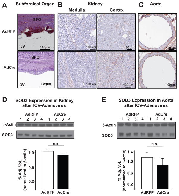
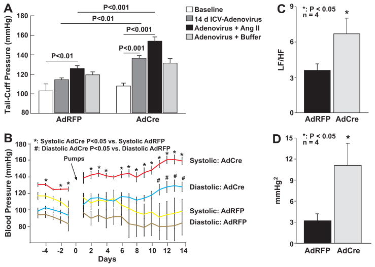
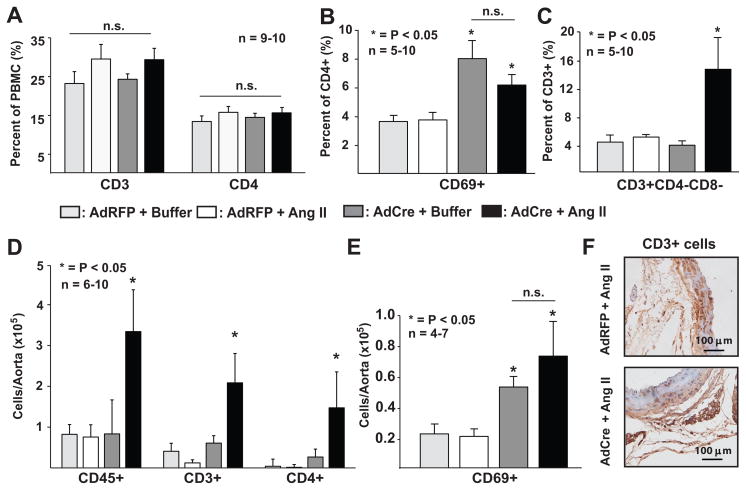
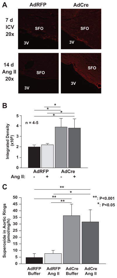
The circumventricular organs (CVOs) lack a well-formed blood-brain barrier and produce superoxide in response to angiotensin II and other hypertensive stimuli. This increase in central superoxide has been implicated in the regulation of blood pressure. The extracellular superoxide dismutase (SOD3) is highly expressed in cells associated with CVOs and particularly with tanycytes lining this region. To understand the role of SOD3 in the CVOs in blood pressure regulation, we performed intracerebroventricular injection an adenovirus encoding Cre-recombinase (5x10(8) particles per milliliter) in mice with loxP sites flanking the SOD3 coding region (SOD3(loxp/loxp) mice). An adenovirus encoding red-fluorescent protein was injected as a control. Deletion of CVO SOD3 increased baseline blood pressure modestly and markedly augmented the hypertensive response to low-dose angiotensin II (140 ng/kg per day), whereas intracerebroventricular injection of adenovirus encoding red-fluorescent protein had minimal effects on these parameters. Adenovirus encoding Cre-recombinase-treated mice exhibited increased sympathetic modulation of heart rate and blood pressure variability, increased vascular superoxide production, and T-cell activation as characterized by increased circulating CD69(+)/CD3(+) cells. Deletion of CVO SOD3 also markedly increased vascular T-cell and leukocyte infiltration caused by angiotensin II. We conclude that SOD3 in the CVO plays a critical role in the regulation of blood pressure, and its loss promotes T-cell activation and vascular inflammation, in part by modulating sympathetic outflow. These findings provide insight into how central signals produce vascular inflammation in response to hypertensive stimuli, such as angiotensin II.
Figures
Comment in
-
Angiotensin II, oxidant signaling, and hypertension: down to a T?Hypertension. 2010 Feb;55(2):228-30. doi: 10.1161/HYPERTENSIONAHA.109.144477. Epub 2009 Dec 14. Hypertension. 2010. PMID: 20008674 Free PMC article. No abstract available.
References
-
- Beyer W, Imlay J, Fridovich I. Superoxide dismutases. Progress in nucleic acid research and molecular biology. 1991;40:221–253. - PubMed
-
- Fukui T, Ishizaka N, Rajagopalan S, Laursen JB, Capers Qt, Taylor WR, Harrison DG, de Leon H, Wilcox JN, Griendling KK. p22phox mRNA expression and NADPH oxidase activity are increased in aortas from hypertensive rats. Circ Res. 1997;80:45–51. - PubMed
-
- Schnackenberg CG, Welch WJ, Wilcox CS. Normalization of blood pressure and renal vascular resistance in SHR with a membrane-permeable superoxide dismutase mimetic: role of nitric oxide. Hypertension. 1998;32:59–64. - PubMed
-
- Jung O, Marklund SL, Geiger H, Pedrazzini T, Busse R, Brandes RP. Extracellular superoxide dismutase is a major determinant of nitric oxide bioavailability: in vivo and ex vivo evidence from ecSOD-deficient mice. Circ Res. 2003;93:622–629. - PubMed
Publication types
MeSH terms
Substances
Grants and funding
LinkOut - more resources
Full Text Sources
Other Literature Sources
Medical
Miscellaneous
