Radiation dose associated with common computed tomography examinations and the associated lifetime attributable risk of cancer
- PMID: 20008690
- PMCID: PMC4635397
- DOI: 10.1001/archinternmed.2009.427
Radiation dose associated with common computed tomography examinations and the associated lifetime attributable risk of cancer
Abstract
Background: Use of computed tomography (CT) for diagnostic evaluation has increased dramatically over the past 2 decades. Even though CT is associated with substantially higher radiation exposure than conventional radiography, typical doses are not known. We sought to estimate the radiation dose associated with common CT studies in clinical practice and quantify the potential cancer risk associated with these examinations.
Methods: We conducted a retrospective cross-sectional study describing radiation dose associated with the 11 most common types of diagnostic CT studies performed on 1119 consecutive adult patients at 4 San Francisco Bay Area institutions in California between January 1 and May 30, 2008. We estimated lifetime attributable risks of cancer by study type from these measured doses.
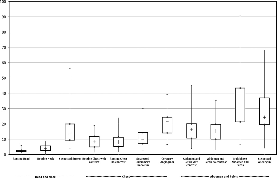
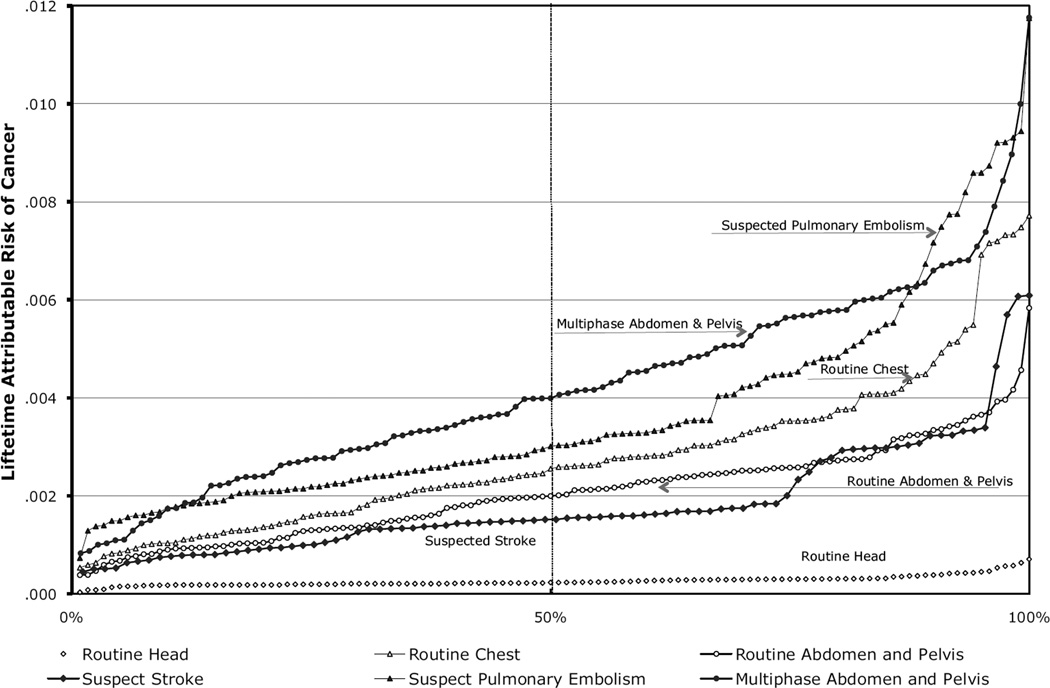
Results: Radiation doses varied significantly between the different types of CT studies. The overall median effective doses ranged from 2 millisieverts (mSv) for a routine head CT scan to 31 mSv for a multiphase abdomen and pelvis CT scan. Within each type of CT study, effective dose varied significantly within and across institutions, with a mean 13-fold variation between the highest and lowest dose for each study type. The estimated number of CT scans that will lead to the development of a cancer varied widely depending on the specific type of CT examination and the patient's age and sex. An estimated 1 in 270 women who underwent CT coronary angiography at age 40 years will develop cancer from that CT scan (1 in 600 men), compared with an estimated 1 in 8100 women who had a routine head CT scan at the same age (1 in 11 080 men). For 20-year-old patients, the risks were approximately doubled, and for 60-year-old patients, they were approximately 50% lower.
Conclusion: Radiation doses from commonly performed diagnostic CT examinations are higher and more variable than generally quoted, highlighting the need for greater standardization across institutions.
Figures
Comment in
-
Cancer risks and radiation exposure from computed tomographic scans: how can we be sure that the benefits outweigh the risks?Arch Intern Med. 2009 Dec 14;169(22):2049-50. doi: 10.1001/archinternmed.2009.453. Arch Intern Med. 2009. PMID: 20008685 No abstract available.
References
-
- Medicare Payment Advisory Commission. [accessed 15 November 2008];A Data Book: Healthcare Spending and the Medicare Program. 2007 Jun; http://www.medpac.gov/documents/Jun07DataBook_Entire_report.pdf.
-
- Amis ES, Jr, Butler PF, Applegate KE, et al. American College of Radiology white paper on radiation dose in medicine. J Am Coll Radiol. 2007 May;4(5):272–284. - PubMed
-
- IMV Medical Information Division. CT census database and market summary report. 2008.
-
- Mettler FA, Jr, Huda W, Yoshizumi TT, Mahesh M. Effective doses in radiology and diagnostic nuclear medicine: a catalog. Radiology. 2008 Jul;248(1):254–263. - PubMed
Publication types
MeSH terms
Grants and funding
LinkOut - more resources
Full Text Sources
Other Literature Sources
Medical
Miscellaneous