. 2009 Dec;127(12):1572-80.
doi: 10.1001/archophthalmol.2009.311.

Melanoma-associated retinopathy: a paraneoplastic autoimmune complication

Affiliations

Melanoma-associated retinopathy: a paraneoplastic autoimmune complication

Ying Lu et al. Arch Ophthalmol. 2009 Dec.

Abstract

Objectives: To study 11 patients with melanoma-associated retinopathy (MAR) to clarify the reliability of various methods of diagnostic testing, to determine the underlying antigenic retinal proteins, and to study the clinical histories and types of associated melanomas.

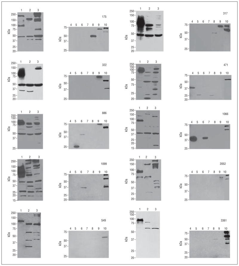
Methods: Clinical data were obtained from patients with melanoma who developed marked visual problems. Testing included electroretinography, kinetic visual fields, comparative studies of Western blots, and indirect immunohistologic examination to detect antiretinal antibodies, as well as proteomic studies to identify underlying antigenic retinal proteins.

Results: Patients with MAR typically have rapid onset of photopsias, scotomata, and loss of central or paracentral vision. Ophthalmoscopy seldom shows significant changes early, but electroretinograms are abnormal. Results of Western blots and immunohistologic examination can show antiretinal antibodies but not always. Most patients (9 of 11) had a strong family history of autoimmune disorders. Any type of melanoma (cutaneous, choroidal, ciliary body, or choroidal nevi) may be associated with this paraneoplastic autoimmune reactivity. MAR may precede or follow the diagnosis of melanoma. Patients with MAR have the same antigenic retinal proteins that have been associated with cancer-associated retinopathy. In addition, 2 new antigenic retinal proteins, aldolase A and aldolase C, were found.

Conclusions: There was a high prevalence of positive family histories of autoimmune disease in patients with MAR. To confirm the disorder, multiple clinical and serum diagnostic techniques (Western blot or indirect immunohistologic examination) are needed. Two newly observed antigenic retinal proteins, aldolase A and aldolase C, are associated with MAR.


Figures

Figure 1
Figure 1
Western blots of serum samples from patients with melanoma-associated retinopathy (patients 175, 317, 322, 471, 886, 1066, 1099, 2052, S49, and 3361) showing immunoreactivity of autoantibodies to (1) human, (2) mouse, and (3) bovine retinal extract. Bands found on human Western blots were isolated and analyzed by mass spectrometry, and results were confirmed by immunoblotting with the following candidate proteins: (4) aldolase A (39 kDa), (5) carbonic anhydrase II (29 kDa), (6) aldolase C (39 kDa), (7) recoverin (23 kDa), (8) S-arrestin (45 kDa), (9) glutathione S-transferase fusion α-enolase (74 kDa), and (10) heat shock protein 60 (60 kDa). Molecular size standards are shown on the left.
Figure 2
Figure 2
Immunostaining analysis of serum from patients with melanoma-associated retinopathy (patients 471, 317, S49, and 175) on human retina, rod outer segments, cone photoreceptor cells, and bipolar cells. Red indicates serum; blue, 4′,6′-diamidino-2-phenylindole nuclear stain; and green, α–protein kinase C (α-PKC) (as a bipolar cell marker). Sample identification numbers correlate with the patients and immunohistologic findings summarized in Table 2. GCL indicates ganglion cell layer; INL, inner nuclear layer; ONL, outer nuclear layer; PRL, photoreceptor layer; and RPE, retinal pigment epithelium. Arrows indicate staining on the retina.
Figure 3
Figure 3
Clinical features of melanoma-associated retinopathy (MAR). A, A 44-year-old woman (patient 2052 in Table 2) had a cutaneous melanoma excised 2 years before developing pericentral scotomata. She was initially seen with generalized retinal atrophy demonstrating no pigment deposits and with rod-cone dysfunction on an electroretinogram (ERG) showing a negative waveform. B, A 73-year-old man (patient S49) had a choroidal melanoma in the right eye requiring enucleation. His remaining eye developed a scotoma 15 months later. Examination showed generalized atrophy of the retina, cone-rod dysfunction on an ERG, and an almost negative waveform. C, A 58-year-old man (patient 322) with a 2-month history of scotomata was found to have a ciliary body mass in the right eye and an overlying conjunctival sentinel vessel. He had generalized depigmented fundus with focal pigment deposits, central scotomata in the right eye, and partial ring scotomata in both eyes. His ERG showed severe cone-rod dysfunction and negative waveforms. The ciliary mass completely regressed over 4 months without treatment, and visual function stabilized in both eyes. D, An 88-year-old man (patient 1066) had progressive deterioration of central and night vision over 10 months, with a large choroidal nevus (7.5-mm wide and 1-mm thick) and surrounding chorioretinal atrophy in the right eye. He had preexisting dry age-related macular degeneration in both eyes with central visual field loss far greater than the area of macular degeneration. Visual fields were asymmetric, with severe loss in the eye with the nevus. The ERG was nonrecordable in the right eye, and his left eye had cone-rod dysfunction with a negative waveform. E, A 40-year-old woman with no ophthalmic disease and with normal retinal appearance underwent visual field tests and an ERG.

References

    1. Sawyer RA, Selhorst JB, Zimmerman LE, Hoyt WF. Blindness caused by photoreceptor degeneration as a remote effect of cancer. Am J Ophthalmol. 1976;81(5):606–613. - PubMed
    1. Suhler EB, Chan CC, Caruso RC, et al. Presumed teratoma-associated paraneoplastic retinopathy. Arch Ophthalmol. 2003;121(1):133–137. - PubMed
    1. Saito W, Kase S, Ohguro H, Furudate N, Ohno S. Slowly progressive cancer-associated retinopathy. Arch Ophthalmol. 2007;125(10):1431–1433. - PubMed
    1. Gass J. Acute Vogt-Koyanagi-Harada-like syndrome occurring in a patient with metastatic cutaneous melanoma. In: Saari K, editor. Uveitis Update: Proceedings of the First International Symposium on Uveitis. Amsterdam, the Netherlands: Elsevier Science; 1984. pp. 407–408.
    1. Keltner JL, Thirkill CE, Yip PT. Clinical and immunologic characteristics of melanoma-associated retinopathy syndrome: eleven new cases and a review of 51 previously published cases. J Neuroophthalmol. 2001;21(3):173–187. - PubMed

Publication types

MeSH terms