. 2009 Dec 15;15(24):7479-7491.
doi: 10.1158/1078-0432.CCR-09-0636.

Resistance to Trastuzumab in Breast Cancer

Affiliations

Resistance to Trastuzumab in Breast Cancer

Paula R Pohlmann et al. Clin Cancer Res. .

Abstract

HER2 is a transmembrane oncoprotein encoded by the HER2/neu gene and is overexpressed in approximately 20 to 25% of invasive breast cancers. It can be therapeutically targeted by trastuzumab, a humanized IgG1 kappa light chain monoclonal antibody. Although trastuzumab is currently considered one of the most effective treatments in oncology, a significant number of patients with HER2-overexpressing breast cancer do not benefit from it. Understanding the mechanisms of action and resistance to trastuzumab is therefore crucial for the development of new therapeutic strategies. This review discusses proposed trastuzumab mode of action as well as proposed mechanisms for resistance. Mechanisms for resistance are grouped into four main categories: (1) obstacles preventing trastuzumab binding to HER2; (2) upregulation of HER2 downstream signaling pathways; (3) signaling through alternate pathways; and (4) failure to trigger an immune-mediated mechanism to destroy tumor cells. These potential mechanisms through which trastuzumab resistance may arise have been used as a guide to develop drugs, presently in clinical trials, to overcome resistance. The mechanisms conferring trastuzumab resistance, when completely understood, will provide insight on how best to treat HER2-overexpressing breast cancer. The understanding of each mechanism of resistance is therefore critical for the educated development of strategies to overcome it, as well as for the development of tools that would allow definitive and efficient patient selection for each therapy. (Clin Cancer Res 2009;15(24):7479-91).


Conflict of interest statement

Disclosure of Potential Conflicts of Interest

The authors have no potential conflicts of interest to disclose.

Figures
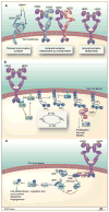
Fig. 1
Fig. 1
HER2 activation. A, receptor dimerization is required for HER2 function (8). In the absence of a ligand, EGFR (represented in blue), HER3, and HER4 assume a tethered conformation. In tethered receptors the dimerization site from extracellular domain II is hidden by intramolecular interactions between domains II and IV. Growth factors alter the conformation of these receptors by binding simultaneously to two sites on extracellular domain domains: I and III (122). HER2 (represented in purple) occurs in an open position and is naturally ready for dimerization. Although no ligand has been identified for HER2 (123), the receptor may become activated by homodimerization (HER2/HER2 pair) or heterodimerization (represented in figure by EGFR/HER2 and HER2/HER3 dimers). The role of HER4 in oncogenesis is less defined (HER2/HER4 dimer not represented). The activation of the HER2 receptor triggers a number of downstream signaling steps through cytoplasm and nucleus, culminating with increased cell growth, survival, and motility (for reviews see refs. , –127). B, activation of PI3K/Akt pathway is one of the most studied processes involved with HER2 activation. PI3K is composed of an 85-kDa regulatory subunit and a 110-kDa catalytic subunit, stably bound to each other and inactive in quiescent cells. Upon activation by the TKs from HER2, p85 binding to the receptor TKs relieves p110α catalytic unit to relocate at the plasma membrane. There p110α phosphorylates and converts phosphatidylinositol (4,5)-bisphosphate (PIP2) into phosphatidylinositol (3,4,5)-triphosphate (PIP3). PIP3 acts as a docking site for pleckstrin homology (PH)-containing proteins, such as Akt and phosphatidylinositol-dependent kinase 1 (PDK1). At the membrane, Akt bound to PIP3 becomes phosphorylated at threonine 308 and serine 473. PDK1 also contributes with Akt activation. Activated Akt activates mTOR and has several other intracellular effects, including interaction with transcription factors, metabolic pathways, apoptosis, and angiogenesis, resulting in cell proliferation, invasion, and survival (128). PIP3 in turn is dephosphorylated back to PIP2 by PTEN. PTEN is therefore a negative regulator of PI3K/Akt signaling and functions as a tumor suppressor. C, the RAS/Raf/MAPK signaling cascade is also triggered by HER2 activation. Growth Factor Receptor-Bound Protein 2 (GRB2) is an adaptor protein that contains one Src Homology 2 (SH2) domain, which recognizes the tyrosine-phosphorylated sites on the activated receptor for binding. GRB2 binds to the guanine nucleotide exchange factor Son of Sevenless (SOS) by one of its SH3 domains. When the GRB2/SOS complex docks to the activated receptor, SOS becomes activated and removes guanosine diphosphate (GDP) from inactive RAS. Free RAS can then bind guanosine-5′-triphosphate (GTP) and become active. RAS/GTP binds efficiently to Raf-1 (MAP3K), which becomes activated. Raf-1 can then activate MEK1 (MAP2K1) and MEK2 (MAP2K2), which are essential signaling nodes downstream RAS and Raf-1. MEK phosphorylates and activates the extracellular signal-regulated kinase (ERK) 1 and ERK2. Activation of ERK results in a broad spectrum of effects relevant to the cell physiology, including cell cycle control, differentiation, and migration, as well as apoptosis and angiogenesis.
Fig. 2
Fig. 2
Trastuzumab schematic structure. The structure of HER2 ectodomain in complex with trastuzumab Fab was described by Cho et al. (23). A, schematic of trastuzumab (IgG1 kappa). Brackets indicate the Fab and the Fc portions of IgG1. CH1 to CH3 indicate the heavy chain constant domains 1 to 3, whereas CL indicates light chain constant domain. VH and VL denote variable heavy chain and variable light chain respectively. B, structural model of human IgG1 VL adapted from Edmundson et al. (129). Complementarity-determining regions 1 to 3 represented in black, yellow, and red are also known as hypervariable regions. Complementarity-determining regions from variable heavy chain and from variable light chains are aligned and form a surface that complements the tridimensional antigen structure. The two sets of six complementarity-determining region loops in the antigen-binding sites are the only murine components of a humanized antibody such as trastuzumab. C, trastuzumab Fab-related function results from its binding to domain IV of HER2. HER2 indicates the human EGFR 2 (in purple). Pertuzumab, another anti-HER2 humanized mAb, binds to an epitope present on domain II of HER2. D, trastuzumab Fc-related functions result from the binding of its Fc portion to other cells that express Fc receptors, such as immune cells, hepatocytes, and endothelial cells. The Fc region of trastuzumab can bind to Fcγ receptor III (RIII) present on the surface of effector cells from the immune system and trigger tumor cell death via ADCC (29). WBC indicates white blood cell.
Fig. 3
Fig. 3
General mechanisms of resistance to trastuzumab: obstacles for trastuzumab binding to HER2. A, a constitutively active truncated form of HER2 receptor that has kinase activity but lacks the extracellular domain and the binding site of trastuzumab is originated from metalloprotease-dependent cleavage of the full-length HER2 receptor (p185; refs. 45, 130). Trastuzumab does not bind to p95HER2 and therefore has no effect against it. The remaining intracellular domain of p95HER2 has operational kinase domains and can be targeted by the TK inhibitor lapatinib (63). B, epitope masking by MUC4 or CD44/polymeric hyaluronan complex. MUC4 is a large membrane-associated mucin produced by epithelia as part of the epithelial protective mechanisms. MUC4 has multiple repeat regions containing serine and threonine. Glycosylation of these repeats forces them into a highly extended, rigid conformation and makes them hydrophilic (69). MUC4 is normally present on the apical surfaces of epithelial cells, but overexpressed in several carcinomas. MUC4 has close association with HER2 and may mask trastuzumab cognate epitope, interfering with antibody binding and activity. CD44/hyaluronan polymer complex activates RAS and PI3K pathways, but it is not clear if these effects depend on HER2. Inhibition of hyaluronan synthesis in vitro reduces hyaluronan polymer binding to CD44, and increases trastuzumab binding to HER2.
Fig. 4
Fig. 4
General mechanisms of resistance to trastuzumab: presence of upregulation of HER2 downstream signaling pathways. PTEN is a tumor suppressor. Trastuzumab binding stabilizes and activates PTEN and consequently down-regulates the PI3K/Akt signaling pathway (39). When PTEN function is lost, PI3K remains constitutively active regardless of binding of trastuzumab to HER2. PTEN loss correlates with clinical unresponsiveness to trastuzumab treatment. Genomic aberrations in the PI3K pathway are a common event in a variety of cancer types (127). Multiple components of this pathway are affected by germline or somatic mutation, amplification, rearrangement, methylation, overexpression, and aberrant splicing, but only in a few instances PIK3CA and PTEN mutations are seen simultaneously (86). Genomic aberrations in the PI3K pathway produce constitutive activation of the pathway, which will signal downstream to the nucleus regardless of trastuzumab binding to HER2. This is the case with activating mutations of PIK3R1 and PIK3CA, encoding genes for PI3K p85α and p110α, respectively. Increased Akt kinase activity and PDPK1 overexpression have also been implicated with trastuzumab resistance.
Fig. 5
Fig. 5
General mechanisms of resistance to trastuzumab: presence of signaling through an alternate receptor and/or pathway. Signaling may continue regardless of trastuzumab binding to HER2 when other receptors remain active on the tumor cell. The activity of certain receptors may also increase as a result of trastuzumab blockage of HER2, as a cell survival mechanism (see text for details). Trastuzumab-induced growth inhibition in HER2-overexpressing cells can be compensated for by increased IGF-IR signaling, resulting in resistance to trastuzumab. In preclinical models in which HER2-overexpressing tumor cells are cultured in the presence of ligand, resembling what is likely to happen in vivo, trastuzumab does not interfere with HER2/HER3 heterodimerization and therefore does not block signaling from these heterodimers (42). c-Met is frequently co-expressed with HER2 in cell lines and contributes to trastuzumab resistance through sustained Akt activation.
Fig. 6
Fig. 6
General mechanisms of resistance to trastuzumab: failure to trigger immune-mediated mechanisms to destroy tumor cells. ADCC is a process in which the Fab region of an antibody binds to its cognate antigen present on a target cell (for instance cancer cell), whereas its Fc region engages with the Fc receptor present on an effector cell from immune system. This process triggers degranulation of cytotoxic granules from effector cell toward the target cell and culminates with target cell apoptosis (131). In humans, the Fc receptor family comprises FcγRI (CD64); FcγRII (CD32), with three isoforms FcγRIIa, FcγRIIb (inhibitory), and FcγRIIc; and FcγRIII (CD16), including two isoforms FcγRIIIa and FcγRIIIb (132). There is a correlation between the clinical efficacy of therapeutic antibodies in humans and their allotype of high-affinity (V158) or low-affinity (F158) polymorphic forms of FcγRIIIa. Epitope masking previously discussed in the obstacles for trastuzumab binding to HER2 section would also play a role by preventing antibody-based cell destruction.

Similar articles

Cited by

References

    1. Perou CM, Sorlie T, Eisen MB, et al. Molecular portraits of human breast tumours. Nature. 2000;406:747–52. - PubMed
    1. Hu Z, Fan C, Oh DS, et al. The molecular portraits of breast tumors are conserved across microarray platforms. BMC Genomics. 2006;7:96. - PMC - PubMed
    1. Sorlie T, Perou CM, Tibshirani R, et al. Gene expression patterns of breast carcinomas distinguish tumor subclasses with clinical implications. Proc Natl Acad Sci U S A. 2001;98:10869–74. - PMC - PubMed
    1. Sorlie T, Tibshirani R, Parker J, et al. Repeated observation of breast tumor subtypes in independent gene expression data sets. Proc Natl Acad Sci U S A. 2003;100:8418–23. - PMC - PubMed
    1. Osborne CK, Yochmowitz MG, Knight WA, 3rd, McGuire WL. The value of estrogen and pro-gesterone receptors in the treatment of breast cancer. Cancer. 1980;46:2884–8. - PubMed