. 2009 Dec 15;23(24):2839-49.
doi: 10.1101/gad.1861409.

miR-19 is a key oncogenic component of mir-17-92

Affiliations

miR-19 is a key oncogenic component of mir-17-92

Virginie Olive et al. Genes Dev. .

Abstract

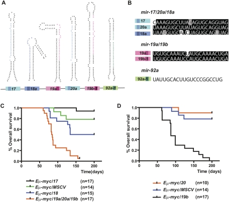
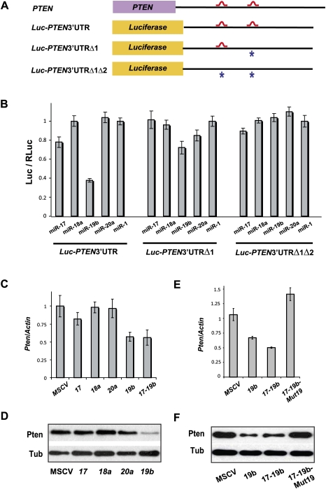
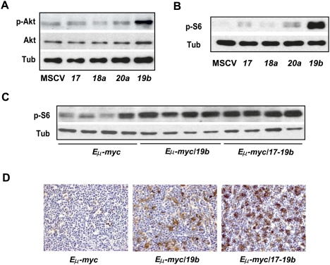
Recent studies have revealed the importance of multiple microRNAs (miRNAs) in promoting tumorigenesis, among which mir-17-92/Oncomir-1 exhibits potent oncogenic activity. Genomic amplification and elevated expression of mir-17-92 occur in several human B-cell lymphomas, and enforced mir-17-92 expression in mice cooperates with c-myc to promote the formation of B-cell lymphomas. Unlike classic protein-coding oncogenes, mir-17-92 has an unconventional gene structure, where one primary transcript yields six individual miRNAs. Here, we functionally dissected the individual components of mir-17-92 by assaying their tumorigenic potential in vivo. Using the Emu-myc model of mouse B-cell lymphoma, we identified miR-19 as the key oncogenic component of mir-17-92, both necessary and sufficient for promoting c-myc-induced lymphomagenesis by repressing apoptosis. The oncogenic activity of miR-19 is at least in part due to its repression of the tumor suppressor Pten. Consistently, miR-19 activates the Akt-mTOR (mammalian target of rapamycin) pathway, thereby functionally antagonizing Pten to promote cell survival. Our findings reveal the essential role of miR-19 in mediating the oncogenic activity of mir-17-92, and implicate the functional diversity of mir-17-92 components as the molecular basis for its pleiotropic effects during tumorigenesis.


Figures

Figure 1.
Figure 1.
miR-19b phenocopies the oncogenic effects of mir-17-92 in the Eμ-myc model. (A) Gene structure of the mir-17-92 polycistronic cluster. (Light-colored boxes) Pre-miRNAs; (dark-colored boxes) mature miRNAs. Homologous miRNA components are indicated by the same or similar colors. (B) mir-17-92 components belong to three miRNA families: miR-17/20a/18 (blue), miR-19 (red), and miR-92a (green). Mature miRNA sequence alignments are shown for each family. Based on sequence identity, miR-17 and miR-20a are closely related homologs, sharing significant sequence identity with miR-18a, but containing a slightly different seed (74% identity among all three). miR-19a and miR-19b differ by a single nucleotide at position 11, and are likely to regulate the same mRNA targets (96% identity). miR-92a has a unique seed sequence that distinguishes it from other components. (C) The mir-19a/20/19b subcluster accelerates c-myc-induced lymphomagenesis. Irradiated mice reconstituted with the Eμ-myc/+ HSPCs overexpressing miR-17, miR-18a, mir-19a/20a/19b, or a MSCV control vector were monitored weekly beginning 4 wk post-transplantation. The Kaplan-Meier curves represent percentage of overall survival. (D) miR-19b accelerates c-myc-induced lymphomagenesis. The mir-19a/20/19b subcluster was further divided into miR-19b and miR-20a, each overexpressed in the Eμ-myc/+ HSPCs before transplantation into lethally irradiated recipient animals. Reconstituted mice were monitored weekly starting 4 wk post-transplantation. The Kaplan-Meier curve indicates miR-19b has a strong oncogenic effect.
Figure 2.
Figure 2.
Eμ-myc/17-19 and Eμ-myc/19b lymphomas have similar pathological and immunological features. (A) Eμ-myc/19b lymphomas are highly invasive. H&E staining of the liver, lung, and kidney showed aggressive invasion by Eμ-myc/19b tumor cells, which was highly analogous to that of the Eμ-myc/19a/20a/19b lymphomas. In particular, both perivascular and parenchymal invasion of the liver were observed. (B) Overexpression of miR-19b represses c-myc-induced apoptosis. The Eμ-myc/19b and Eμ-myc/19a/20a/19b lymphomas had similar proliferation rates to those of Eμ-myc/MSCV controls, demonstrated by Ki67 staining. However, exogenous expression of miR-19b or mir-19a/20/19b greatly decreased apoptosis in the Eμ-myc tumors, confirmed by TUNEL and H&E staining of Eμ-myc/17-19 lymph node tumors. The “starry sky” morphology of cell clusters undergoing apoptosis (black arrows), a hallmark of Eμ-myc/MSCV lymphomas, was absent in Eμ-myc/19b and Eμ-myc/19a/20a/19b tumors. (C) miR-19b and mir-17-19b preferably transform progenitor B cells. Flow cytometric immunophenotyping of representative Eμ-myc, Eμ-myc/17-19, and Eμ-myc/19 lymphomas. While the majority of Eμ-myc tumors consisted of CD19-positive and IgM-positive B cells, the Eμ-myc/19b and Eμ-myc/17-19 tumor cells bore cellular characteristics of progenitor B cells, positive for CD19 but not for surface IgM.
Figure 3.
Figure 3.
miR-19 miRNAs are essential for the oncogenic activity of mir-17-19b. (A) A schematic representation of the gene structural organization of mir-17-19b, mir-17-19b-Mut19, and miR-19b. 19a* and 19b* indicate miR-19 mutations. (B) miR-19 mutations specifically affected miR-19 expression in mir-17-19b. 3T3 cells were infected with MSCV-mir-17-19b, MSCV-mir-17-19b-Mut19, or control MSCV vectors. Expression levels of miR-17, miR-18a, miR-19a, miR-20a, and miR-19b were determined using TaqMan miRNA assays. miR-19 mutations specifically affected the expression of miR-19a and miR-19b, but not that of the adjacent mir-17-19b components. Error bars indicate SD (n = 3). (C) miR-19 is both necessary and sufficient for the oncogenic effect of mir-17-19b. Overexpression of miR-19b and mir-17-19b accelerated c-myc-induced lymphomagenesis to a similar degree, shown in Kaplan-Meier curves as the percentage of overall survival. Mutations in miR-19 greatly decreased the oncogenic activity of mir-17-19b in the Eμ-myc model. We compared the oncogenic effects of mir-17-19b, miR-19b, and mir-17-19b-Mut19 in the same adoptive transfer experiment. (D) miR-19 is both necessary and sufficient for the cell survival effect of mir-17-19b in vivo. Representative lymphomas from Eμ-myc, Eμ-myc/19b, and Eμ-myc/17-19b-Mut19 were stained for H&E and caspase-3, which indicated that the miR-19 mutations significantly compromised cell survival effects of mir-17-19b, while miR-19b overexrepssion suppresses apoptosis. The Eμ-myc/19b and Eμ-myc/17-19b-Mut19 tumors shown here were both IgM-negative B lymphomas. (Arrow) “Starry sky” feature of apoptotic tumor cells; (arrowhead) apoptotic cells with positive caspase-3 staining. Bar, 50 μm. (E) Quantification of caspase-3 staining of representative tumors from D as percentage of positive cells. For Eμ-myc/19b and Eμ-myc/17-19b-Mut19 tumors, only IgM-negative tumors were selected for this comparison (n = 3; error bars represent SEM).
Figure 4.
Figure 4.
Pten is a mir-17-19b target specifically regulated by miR-19. (A) Schematic representation of the PTEN 3′UTR and its miR-19-binding sites. Two miR-19-binding sites (shown in red) can be found in the human PTEN 3′UTR. The PTEN 3′UTR with mutations (designated with asterisks) at one (PTEN3′UTRΔ1) or both (PTEN3′UTRΔ1Δ2) miR-19 sites, as well as the wild-type counterpart (PTEN3′UTR), were each cloned downstream from a luciferase reporter (Luc). (B) Specific repression of Luc-PTEN3′UTR reporter by miR-19. Luc-PTEN3′UTR was cotransfected with mimics of miR-17, miR-18a, miR-19b, miR-20a, and control miR-1. Only miR-19b significantly repressed the reporter expression. Cotransfection with a luciferase construct carrying one mutated miR-19b site in the PTEN 3′UTR (Luc-PTEN3′UTRΔ1) partially derepressed the Luc reporter, and cotransfection of a construct with mutations in both miR-19 sites (Luc-PTEN3′UTRΔ1Δ2) completely derepressed the Luc reporter. (C,D) miR-19b specifically represses endogenous Pten expression level. Using real-time PCR analysis (C) and Western analysis (D), down-regulation of endogenous Pten mRNA and protein can be detected in serum-starved NIH-3T3 cells infected with miR-19b and mir-17-19b. In comparison, overexpression of miR-17, miR-18a, miR-20a, and control vector (MSCV) in these cells has minimal effects on the endogenous Pten level. (E,F) miR-19 is both necessary and sufficient to mediate the Pten repression by mir-17-19b. Repression of endogenous Pten mRNA (E) and protein (F) can be detected in serum-starved NIH-3T3 cells infected with miR-19b and mir-17-19b. In comparison, mir-17-19b-Mut19 failed to repress the endogenous Pten level.
Figure 5.
Figure 5.
miR-19b functionally antagonizes pten-induced apoptosis in Xenopus embryos. (A,B) miR-19b rescues hydroxyurea (HU)-induced apoptosis in X. laevis embryos. Injection of miR-19b mimics into X. laevis embryos rescued apoptosis caused by hydroxyurea treatment. The mutated miR-19b with an altered seed region (mut-19b) failed to rescue hydroxyurea-induced apoptosis. Embryos undergoing apoptosis were marked by cell blebbing, disruption of cell adhesion, and a characteristic white color (red arrowhead). Hydroxyurea-treated embryos appeared more pigmented than control embryos, largely due to developmental arrest. (B) Apoptosis was quantified for untreated and hydroxyurea-treated embryos, as well as the hydroxyurea-treated embryos coinjected with either miR-19b mimics or mut-19b mimics (n = 3 experiments with >25 embryos in each group; [*] P < 0.05). (C,D) Injection of miR-19b rescued pten-induced apoptosis in Xenopus embryos. Injection of full-length pten mRNA into Xenopus embryos led to widespread apoptosis. Injection of miR-19b, but not mut-19b, significantly rescued the proapoptotic effect of pten. (E,F) Disruption of base-pairing between miR-19 and pten mRNA abolished their functional antagonism. Mutations in the miR-19-binding sites within the pten mRNA (pten-mut) did not abrogate the proapoptotic effects of pten, but did eliminate the ability of miR-19b to repress the apoptosis (n = 3 experiments with >25 embryos in each group; [*] P < 0.05). All error bars represent SEM.
Figure 6.
Figure 6.
miR-19 and mir-17-19b activates the Akt–mTOR pathway. (A) miR-19 is a key mir-17-19b component to activate the Akt–mTOR pathway. Using Western analysis, increased phospho-Akt level was detected in serum-starved 3T3 cells infected with miR-19b, but not miR-17, miR-18a, miR-20a, and the control vector (MSCV). In comparison, the overall Akt level was not affected by miR-19b. (B) miR-19 induces an increase in phosphorylation of S6 ribosomal protein. Enforced expression of miR-19b strongly promoted the S6 phosphorylation as compared with the rest of mir-17-19b components. (C,D) Enforced miR-19b or mir-17-19b expression in the Eμ-myc model led to an increased level of phospho-S6 in lymphomas. Cells derived from the Eμ-myc, Eμ-myc/19b, and Eμ-myc/17-19b lymphomas were analyzed by Western (C) and immunohistochemistry (D). Both Eμ-myc/19b and Eμ-myc/17-19b lymphomas exhibited a high level of phospho-S6, although mir-17-19b seemed to have a stronger effect. In comparison, Eμ-myc tumors exhibited a low level of phospho-S6 and more variation among different samples, possibly reflecting the differences in the secondary oncogenic lesions. In all Western analyses, tubulin (Tub) was used as a normalization control.

Comment in

Similar articles

Cited by

References

    1. Abubaker J, Bavi PP, Al-Harbi S, Siraj AK, Al-Dayel F, Uddin S, Al-Kuraya K. PIK3CA mutations are mutually exclusive with PTEN loss in diffuse large B-cell lymphoma. Leukemia. 2007;21:2368–2370. - PubMed
    1. Adams JM, Harris AW, Pinkert CA, Corcoran LM, Alexander WS, Cory S, Palmiter RD, Brinster RL. The c-myc oncogene driven by immunoglobulin enhancers induces lymphoid malignancy in transgenic mice. Nature. 1985;318:533–538. - PubMed
    1. Ambros V. The functions of animal microRNAs. Nature. 2004;431:350–355. - PubMed
    1. Baek D, Villen J, Shin C, Camargo FD, Gygi SP, Bartel DP. The impact of microRNAs on protein output. Nature. 2008;455:64–71. - PMC - PubMed
    1. Bartel DP. MicroRNAs: Target recognition and regulatory functions. Cell. 2009;136:215–233. - PMC - PubMed

Publication types