. 2009 Dec 15;23(24):2876-86.
doi: 10.1101/gad.1863009.

Nearby inverted repeats fuse to generate acentric and dicentric palindromic chromosomes by a replication template exchange mechanism

Affiliations

Nearby inverted repeats fuse to generate acentric and dicentric palindromic chromosomes by a replication template exchange mechanism

Ken'Ichi Mizuno et al. Genes Dev. .

Abstract

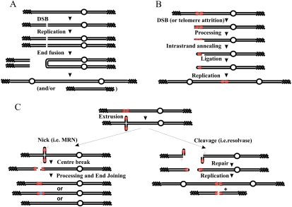
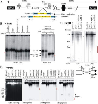
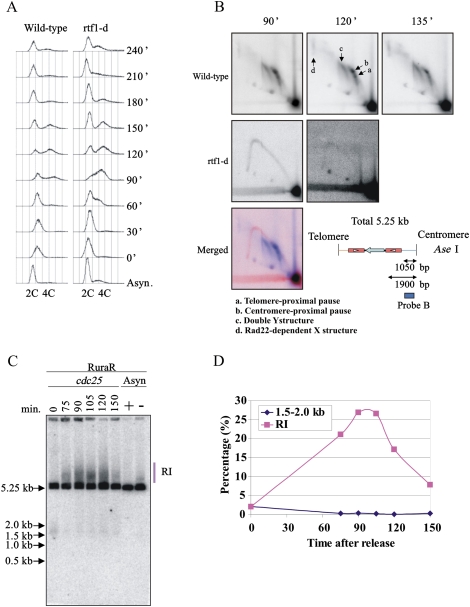
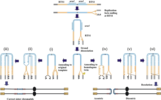
Gene amplification plays important roles in the progression of cancer and contributes to acquired drug resistance during treatment. Amplification can initiate via dicentric palindromic chromosome production and subsequent breakage-fusion-bridge cycles. Here we show that, in fission yeast, acentric and dicentric palindromic chromosomes form by homologous recombination protein-dependent fusion of nearby inverted repeats, and that these fusions occur frequently when replication forks arrest within the inverted repeats. Genetic and molecular analyses suggest that these acentric and dicentric palindromic chromosomes arise not by previously described mechanisms, but by a replication template exchange mechanism that does not involve a DNA double-strand break. We thus propose an alternative mechanism for the generation of palindromic chromosomes dependent on replication fork arrest at closely spaced inverted repeats.


Figures

Figure 1.
Figure 1.
Generation of palindromic chromosomes. (A) An uncapped end is generated by a DSB (or by telomere attrition, not shown). The resulting molecule is then replicated, producing two identical uncapped sister chromatids. The two free ends are directly ligated (for example, by NHEJ), and this results in a palindromic dicentric or acentric palindrome. Open circles indicate centromeres, zig-zag lines represents telomeres, and single lines indicate ssDNA. (B) An uncapped end near a short inverted repeat (in red) can, after processing to reveal ssDNA, result in a “snap-back” strand annealing between the inverted repeats, generating a hairpin at the chromosome end. Following fill-in and ligation, this will generate a capped end that resolves into a giant palindrome after DNA replication. (C) If the palindrome extrudes, it will generate a cruciform structure. This resembles an HJ, has hairpin ends, and can be processed by a structure-specific nuclease such as Mus81 (right) or by the MRN complex (left). (Left) If both hairpins are cleaved at the apex by MRN, this results in a break at the center of the palindrome. This can subsequently be repaired by end-joining. This process can result in gain or loss of sequences at the center of the palindrome (blue), and ultimately result in its stabilization or complete loss. If the extrusion is cleaved by HJ resolvase, it is able to form hairpin-capped chromosomes by ligation. Following DNA replication, these can result in acentric and dicentric giant palindromes. This process is suppressed by MRN activity, which can open the hairpin at the chromosome end, thus generating an uncapped end, a substrate for schematic A.
Figure 2.
Figure 2.
Replication fork arrest within a palindrome causes the formation of acentric and dicentric chromosomes. (A) Schematic of the constructs integrated at the ura4 locus on ChrIII. (Blue) RTS1; polar RTS1. The bar in RTS1 indicates the direction that, when a fork approaches, it will be arrested. Red arrows below the ura4 (yellow) and his3 (green) sequences indicate the direction of transcription. Boxes Cen and Tel indicate position of probes used. Ovals show the ars sequences. (Inset) Schematic of acentric and dicentric chromosomes. The 14-bp insertion interrupts the symmetry of the palindrome center in RuiuR and oRuiuRo. (B) Viability analysis of perfect (RuuR) and interrupted (RuiuR) palindrome strains in the presence (pause on; +, rtf1+) and absence (pause off; −, rtf1+ or rtf1-d) of fork arrest. Note that loss of viability is dependent on fork arrest by rtf1+ induction. (C) Representative images of RuiuR mitotic catastrophe 24 h after thiamine removal (approximately three generations of growth under conditions in which forks arrest at RTS1). DNA stained with DAPI, and septum stained with Calcofluor. (D) PFGE and Southern blot analysis of strains either unable to arrest forks at RTS1 (rtf1Δ) or where rtf1 is regulated by thiamine ([−] low transcript and minimal arrest; [+] high transcript and efficient fork arrest). Note that formation of 1.4-Mb DNA is correlated with loss of viability in B. (E) PFGE and Southern blot analysis of cells without fork arrest (off; −) and 24 h after (on; +) arrest is induced. (Left panel) Ethidium bromide staining. (Middle panel) Telomere-proximal probe. (Right panel) Centromere-proximal probe. I, II and III indicate fission yeast chromosomes. Arrows: 3.5 Mb, ChrIII; 1.4 Mb, acentric; 5.6 Mb, dicentric. (*) Smear corresponding to breakage.
Figure 3.
Figure 3.
Fork arrest at inverted repeats also results in large palindromic chromosomes. (A) PFGE of DNA from the indicated strains either without (off; −) or 48 h after (on; +) the induction of rtf1-dependent fork arrest. (Left) Ethidium stained. (Right) Southern blotted with a telomere-proximal probe. The percentage of signal corresponding to the acentric chromosome (1.4 Mb) was calculated (numerals) as a percentage of total signal (2 × 3.5 Mb + 1.4 Mb). (B, left) Schematic. (Blue) RTS1. (Bar in RTS1) The direction that, when a fork approaches, it will be arrested. Red arrows below ura4 (yellow) and his3 (green) indicate direction of transcription. (Solid triangle) 14-bp “interruption”. (Right) Viability analysis in the absence (Pause on) or presence (Pause off) of thiamine to regulate rtf1 for the strains indicated. (C) Schematic of the expected pattern of replication arrest for RuiuR and oRuiuRo. The blue bar indicates the direction that, when a fork approaches, it will be arrested.
Figure 4.
Figure 4.
HR proteins are required for viability and GCR. (A,B) DNA prepared from RuiuR strains indicated, grown either without (off; −) or three generations after (on; +) the induction of rtf1-dependent fork arrest, was analyzed by either PFGE (left) or SalI digestion (right) (see Supplemental Fig. S3C for SalI fragments) and Southern blotting with a telomere-proximal probe. The panel labeled “rad60” represents the same gel probed with a ChrII-specific probe. (Bottom) The ratio of signal corresponding to the acentric chromosome (1.4 Mb or 10.3 kb, respectively, expressed as a percentage) was calculated compared with total signal (2 × 3.5 Mb + 1.4 Mb or 2 × 19.8 kb + 10.3 kb). The rad22Δ rhp51Δ strain was used because rad22rad52 mutants can accumulate suppressors that are Rhp51Rad51-dependent. rad32 = mre11. (C) Viability analysis in the presence (Pause off) or absence (Pause on) of thiamine to regulate rtf1.
Figure 5.
Figure 5.
Fork arrest at RuiuR or RuraR results in chromosomal rearrangement without DSB formation in rad+ cells and in processing (rad50) and recombination (rad22rad52) of defective mutants. (A) Schematic of the RuiuR or RuraR constructs integrated at the ura4 locus of ChrIII. The bar in RTS1 indicates the direction that, when a fork approaches, it will be arrested. Boxes ade6 and rng3, the bar cen, and the circle cent3 indicate the positions of probes. Note that ARS3004 and ARS3005 are efficient ARSs. The single AscI site is indicated. (B, left) Southern hybridization of a PFG using a centromere-proximal probe either without the induction of rtf1-dependent replication fork arrest (pause −) or 48 h after the induction (pause +) in the indicated RuiuR strains. If a DSB occurred at RuiuR, a 2.8-Mb band would represent a fragment derived from the DSB to the right telomere. This was not seen; the expected position is indicated by a red bar. (Right) AscI-digested DNA was analyzed by PFGE and Southern blotting with a centromere-proximal probe. The AscI site is a unique site on S. pombe ChrIII and would generate a 650-kb fragment if combined with a DSB at RuiuR. No such signal is seen; compare tracks 3 and 4. The expected position is indicated by a red bar. (C) Southern blot analysis of AvaI restriction fragment derived from indicated RuraR strains without (−) and 24 h after (+) arrest is induced. Asterisk (*) represents a nonspecific band. Below is a schematic of the RuraR construct digested by the restriction enzyme AvaI. A polar DSB would result in the generation of a 2.2-kb fragment revealed with the Cen probe. This was not seen; the expected position is indicated by a red bar. (D, left) A PFG of chromosomes from the indicated RuraR strains stained with ethidium bromide or probed with indicated probes. (−) Pause off; (+) pause on for 24 h. Positions of H. wingei chromosomes are indicated as size markers. The bold arrow indicates the position of the acentric chromosome at 1.4 Mb. The predicted fragment in the case of a DSB at RuraR is 0.7 Mb for the Rng3 probe. The ade6 and cent3 probes would show a 2.8-Mb fragment in the case of a DSB. These were not detected; the expected positions are indicated on the right by a red bar. In the absence of both Rad50 and Rad22Rad52 (DSB resection and HR), a smear above 2.8 Mb is evident. However, this appeared whatever ChrIII probe was used, which thus does not reflect a specific polar DSB, but likely reflects fragmentation of the chromosome.
Figure 6.
Figure 6.
Absence of a detectable DSB induction in synchronized cells. RuraR rtf1+ and RuraR rtf1-d cells were grown without thiamine (14 h) and were synchronized by cdc25.22 block and release. The time course starts after cell cycle release. Asynchronous cultures were grown for 24 h with (−; pause off) and without (+; pause on) thiamine as a control where appropriate. (A) FACS analysis. The culture before the cell cycle arrest was used as asynchronous control culture “Async.” S phase occurs between 60 and 150 min. (B) Two-dimensional gel electrophoresis at specified time points with (rtf1+) and without (rtf1-d) fork arrest (a–d as indicated) (see Supplemental Fig. S1). (Bottom) Map of AseI digest for RuraR. The position of the Cen probe is indicated by the bar. In the merged picture, the signal corresponding to 90 min for rtf1-d is red and that for rtf1+ is blue. (C) Southern hybridization of one-dimensional gel to visualize replication intermediates (RI) from the indicated times. DNA was digested with AseI. (D) Quantification of one-dimensional gel showing the percentage total signal present in replication intermediates (red squares) and at the position of a predicted break (blue diamonds). Forks broken within the Cen-proximal RTS1 would generate a band of ∼1500 bp. A faint signal between 1.5 and 2.0 kb is seen in the G2-arrested sample (0 min), but this is lost when cells enter S phase (75 min).
Figure 7.
Figure 7.
An RTE model for giant palindrome formation. After fork arrest, a recombinogenic 3′ end is formed by association with HR proteins (yellow). (Left) Recombination protein-dependent fork restart results in reinvasion at the correct locus (i) and completion of replication (ii,iii). (Right, iv) Alternatively, erroneous invasion occurs in the opposite repeat. The dashed line indicates an area synthesised by restart of coupled leading and lagging strand synthesis. Replication subsequently continues around the palindrome (v) and creates an HJ following ligation to the lagging strand of the oncoming fork (vi). HJ resolution in the horizontal plane results in acentric and dicentric chromosome formation. Resolution in the vertical plane results in fully replicated chromosomes in the original conformation (not shown).

Comment in

  • Leaping forks at inverted repeats.
    Branzei D, Foiani M. Branzei D, et al. Genes Dev. 2010 Jan 1;24(1):5-9. doi: 10.1101/gad.1884810. Genes Dev. 2010. PMID: 20047996 Free PMC article.

Similar articles

Cited by

References

    1. Aguilera A, Gomez-Gonzalez B. Genome instability: A mechanistic view of its causes and consequences. Nat Rev Genet. 2008;9:204–217. - PubMed
    1. Albrecht EB, Hunyady AB, Stark GR, Patterson TE. Mechanisms of sod2 gene amplification in Schizosaccharomyces pombe. Mol Biol Cell. 2000;11:873–886. - PMC - PubMed
    1. Bahler J, Wu JQ, Longtine MS, Shah NG, McKenzie A, 3rd, Steever AB, Wach A, Philippsen P, Pringle JR. Heterologous modules for efficient and versatile PCR-based gene targeting in Schizosaccharomyces pombe. Yeast. 1998;14:943–951. - PubMed
    1. Bailey JA, Gu Z, Clark RA, Reinert K, Samonte RV, Schwartz S, Adams MD, Myers EW, Li PW, Eichler EE. Recent segmental duplications in the human genome. Science. 2002;297:1003–1007. - PubMed
    1. Bartkova J, Horejsi Z, Koed K, Kramer A, Tort F, Zieger K, Guldberg P, Sehested M, Nesland JM, Lukas C, et al. DNA damage response as a candidate anti-cancer barrier in early human tumorigenesis. Nature. 2005;434:864–870. - PubMed

Publication types

MeSH terms

LinkOut - more resources