. 2009 Dec 15;23(24):2915-24.
doi: 10.1101/gad.544009.

Human POT1 is required for efficient telomere C-rich strand replication in the absence of WRN

Affiliations

Human POT1 is required for efficient telomere C-rich strand replication in the absence of WRN

Nausica Arnoult et al. Genes Dev. .

Abstract

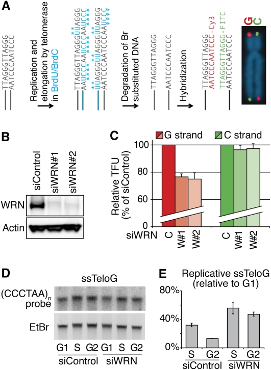
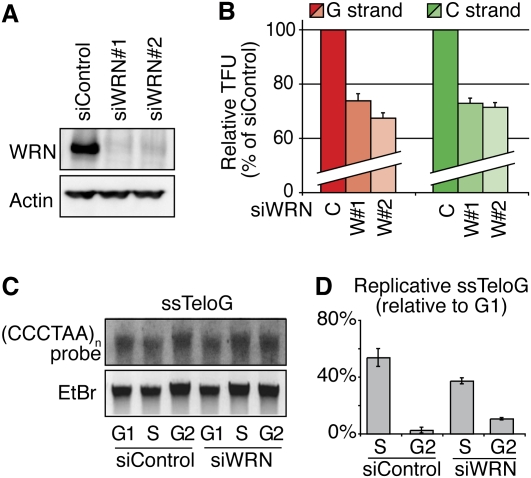
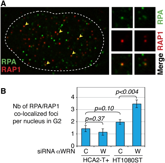
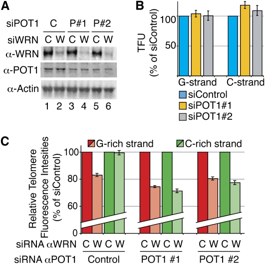
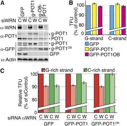
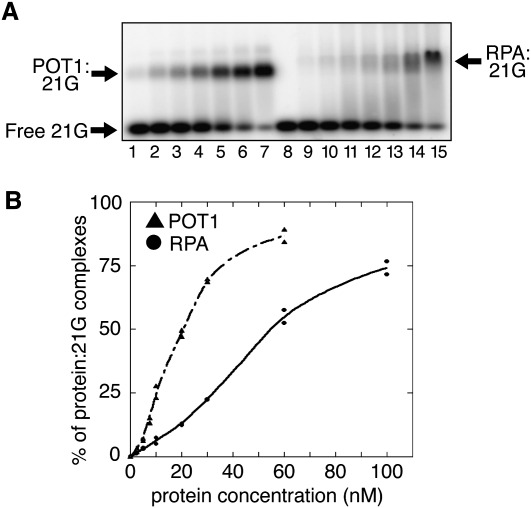
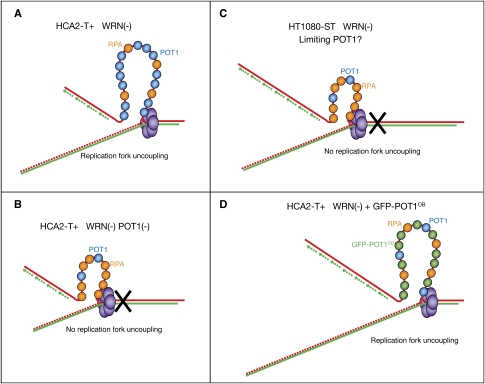
Mechanisms of telomere replication remain poorly defined. It has been suggested that G-rich telomeric strand replication by lagging mechanisms requires, in a stochastic way, the WRN protein. Here we show that this requirement is more systematic than previously thought. Our data are compatible with a situation in which, in the absence of WRN, DNA synthesis at replication forks is uncoupled, thus allowing replication to continue on the C strand, while single G strands accumulate. We also show that in cells in which both WRN and POT1 are limiting, both G- and C-rich telomeric strands shorten, suggesting a complete replication block. Under this particular condition, expression of a fragment spanning the two POT1-OB (oligonucleotide-binding) fold domains is able to restore C (but not G) strand replication, suggesting that binding of POT1 to the lagging strand allows DNA synthesis uncoupling in the absence of WRN. Furthermore, in vitro experiments indicate that purified POT1 has a higher affinity for the telomeric G-rich strand than purified RPA. We propose a model in which the relative enrichments of POT1 versus RPA on the telomeric lagging strand allows or does not allow uncoupling of DNA synthesis at the replication fork. Our study reveals an unanticipated role for hPOT1 during telomere replication.


Figures

Figure 1.
Figure 1.
Depletion of WRN leads to replication fork uncoupling at telomeres. (A) In the CO-FISH technique, BrdU and BrdC are incorporated during S phase. Br-substituted strands (i.e., neosynthesized by replication or telomerase elongation) are degraded by the CO-FISH procedure. Fluorescence intensities of hybridized probes revealing the telomeric G- and C-rich strands reflect the length of parental strands after replication. (B) Efficient knockdown (95%) of WRN in HCA2-T+ cells after transfection of siRNAs and revealed by Western blot. (C) WRN knockdown in these cells leads to G-rich strand shortening (red), while C-rich strands (green) are completely replicated. Average measurements of 30 metaphases are represented as percentages (%) of control siRNA (single intensity distributions are presented in Supplemental Fig. S1). Partial WRN knockdown (<90%) allows normal telomere replication (not shown). (D) Accumulation of replicative single G strands after WRN knockdown shown by the telomeric single-strand assay (Gomez et al. 2004). Radioactive signals are normalized with regard to total DNA input (ethidium bromide). The signal corresponding to single G strands detected in cells blocked in G1 phase is considered as the baseline. (E) Quantification of single G strand signals shows that this strand accumulates at higher levels during S phase in the absence of WRN and that it persists in G2.
Figure 2.
Figure 2.
No replication fork uncoupling after WRN knockdown in cells with very long telomeres. (A) Efficient knockdown of WRN in HT1080-ST cells revealed by Western blot. (B) In ST cells, WRN knockdown leads to both C-rich strand (green) and G-rich strand shortening (red). Average measurements of 30 metaphases, represented as percentages (%) of control siRNA (single intensity distributions are presented in Supplemental Fig. S4). (C) Accumulation of replicative single G strands after WRN knockdown shown by the telomeric single-strand assay. Radioactive signals are normalized with regard to total DNA input (ethidium bromide). The signal corresponding to single G strands detected in cells blocked in G1 phase is considered as the baseline. (D) Quantification of single G strands indicates that ST cells deficient in WRN do not accumulate more replicative single G strands during S phase and that these strands do not persist in G2.
Figure 3.
Figure 3.
Higher RPA accumulation at telomeres of ST cells upon depletion of WRN. (A) Example of colocalized RPA (green) and telomere (RAP1, red) foci revealed by deconvolution three-dimensional IF after cell sorting for cells in G2 phase. (B) Quantification of RPA/RAP1 colocalization foci. The number of RPA/RAP1 foci after WRN knockdown in G2 in normal fibroblasts (HCA2-T+) remains the same, while there is a small but highly significant increase of this number in ST cells depleted for WRN. At least 30 nuclei were analyzed.
Figure 4.
Figure 4.
POT1 partial knockdown prevents replication fork uncoupling at telomeres of WRN-depleted HCA2-T+ cells. (A) Efficient knockdown of WRN (>90%) (lanes 2,4,6) combined to partial knockdown of POT1 (50%–60% compared with control) (lanes 1,2) with two different POT1 siRNAs (P#1, lanes 3,4; P#2, lanes 5,6) in HCA2-T+, revealed by Western blot. (B) POT1 partial knockdowns have no effect on telomere replication. The histogram represents mean CO-FISH signal intensities after POT1 knockdown, represented as percentages (%) of control siRNA. (C) POT1 knockdown leads to shortening of both C-rich (green) and G-rich (red) strands in HCA2-T+ cells depleted for WRN. Quantification results are represented in percentages of control siRNA for WRN.
Figure 5.
Figure 5.
Forced expression of GFP-POT1-OB1+2 in ST cells depleted for WRN rescues replication fork uncoupling at telomeres. (A) Efficient knockdown of WRN (>90%) combined with forced expression of GFP-POT1(fl) or GFP-POT1-OB1+2. POT1 antibodies reveal both endogenous POT1 and GFP-POT1(fl) (g-POT1). GFP antibodies reveal proteins expressed from all three transient transfection experiments: control (GFP), GFP- POT1(fl) (g-POT1), and GFP-POT1-OB1+2 (g-POT1OB). (B) Neither GFP-POT1(fl) nor GFP-POT1-OB1+2 expressions by themselves affect parental telomere length after replication. CO-FISH signal quantifications after POT1 knockdown are represented in percentages (%) of control transfected with the GFP plasmid. (C) Expression of GFP-POT1-OB1+2 in ST cells depleted for WRN is able to completely rescue C strand (but not G strand) replication, while forced expression of GFP-POT1(fl) has no impact. Quantification results are represented in percentages of control siRNA for WRN.
Figure 6.
Figure 6.
POT1 binds to telomeric G-rich single strands with higher affinity than RPA. (A) Titration of 21G as a function of hPOT1 or hRPA concentration. 32P-21G (20 nM) (lane 8: without protein) was incubated with different amounts of POT1 (lane 1: 2.5 nM; lane 2: 5 nM; lane 3: 7.5 nM; lane 4: 10 nM; lane 5: 20 nM; lane 6: 30 nM; lane 7: 60 nM) or RPA (lane 9: 2.5 nM; lane 10: 5 nM; lane 11: 10 nM; lane 12: 20 nM; lane 13: 30 nM; lane 14: 60 nM; lane 15: 100 nM) and separated on a 1% agarose gel. (POT1:21G) Noncovalent complexes between 21G and POT1; (RPA:21G) noncovalent complexes between 21G and RPA. (B) Quantitation of POT1 and RPA titrations. Each band was analyzed and quantified with a PhosphorImager Storm 860 instrument (Molecular Dynamics). The data from two independent experiments are shown. POT1 binding is represented by triangles connected by a dashed line, and RPA binding is represented by circles connected by a solid line.
Figure 7.
Figure 7.
Model for replication fork uncoupling upon WRN loss. (A) In normal fibroblasts (HCA2-T+), the single G strands that accumulate during replication are bound by POT1, thus preventing accumulation of RPA and fork stalling. This accumulation therefore enables polymerization to continue on the leading strands up to the end of the chromosome. (B) When combined with POT1 deficiency, WRN depletion in HCA2-T+ cells leads to RPA accumulation on single G strands, thus inducing replication fork stall. (C) In ST cells, levels of POT1 are limiting, but enough to ensure normal telomere replication. After WRN knockdown, there is a deficit of POT1 molecules so that RPA accumulates on single G strands, thus preventing replication fork uncoupling, leading to both G-rich and C-rich strand shortening. (D) Forced expression of the OB fold domains of POT1 prevents RPA from binding to single G strands, thereby restoring the replication fork uncoupling mechanism, which allows complete C strand replication.

References

    1. Alani E, Thresher R, Griffith JD, Kolodner RD. Characterization of DNA-binding and strand-exchange stimulation properties of y-RPA, a yeast single-strand-DNA-binding protein. J Mol Biol. 1992;227:54–71. - PubMed
    1. Arnoult N, Shin-Ya K, Londoño-Vallejo JA. Studying telomere replication by Q-CO-FISH: The effect of telomestatin, a potent G-quadruplex ligand. Cytogenet Genome Res. 2008;122:229–236. - PubMed
    1. Bae SH, Bae KH, Kim JA, Seo YS. RPA governs endonuclease switching during processing of Okazaki fragments in eukaryotes. Nature. 2001;412:456–461. - PubMed
    1. Baird DM, Davis T, Rowson J, Jones CJ, Kipling D. Normal telomere erosion rates at the single cell level in Werner syndrome fibroblast cells. Hum Mol Genet. 2004;13:1515–1524. - PubMed
    1. Barrientos KS, Kendellen MF, Freibaum BD, Armbruster BN, Etheridge KT, Counter CM. Distinct functions of POT1 at telomeres. Mol Cell Biol. 2008;28:5251–5264. - PMC - PubMed

Publication types

LinkOut - more resources