. 2009 Dec;2(6):611-9.
doi: 10.1161/CIRCEP.109.872093.

Catheter ablation of atrial fibrillation without fluoroscopy using intracardiac echocardiography and electroanatomic mapping

Affiliations

Catheter ablation of atrial fibrillation without fluoroscopy using intracardiac echocardiography and electroanatomic mapping

John D Ferguson et al. Circ Arrhythm Electrophysiol. 2009 Dec.

Abstract

Background: Catheter ablation of atrial fibrillation is currently guided by x-ray fluoroscopy. The associated radiation risk to patients and medical staff may be significant. We report an atrial fibrillation ablation technique using intracardiac echocardiography (ICE) and electroanatomic mapping without fluoroscopy.

Methods and results: Twenty-one patients with atrial fibrillation (age, 42 to 73 years; 14 male; 14 paroxysmal, 7 persistent; body mass index, 26 to 38) underwent ablation. A decapolar catheter was advanced through the left subclavian vein until stable coronary sinus electrograms appeared on all electrodes. Two 9F sheaths were advanced transfemorally over a guide wire to the right atrium. A rotational ICE catheter was advanced through a deflectable sheath. Double transseptal puncture was performed with ICE guidance and facilitated by electrocautery. A 3D MRI left atrial image was registered to the ostia of the pulmonary veins using ICE. Catheter ablation was performed using ICE and electroanatomic mapping navigation. In 19 cases, no fluoroscopy was used and the staff did not wear protective lead. In 2 cases, 2 to 16 minutes of fluoroscopy was used to assist transseptal puncture. Median procedure time was 208 (188 to 221) minutes; coronary sinus cannulation took 5 (2 to 26) minutes; double transseptal took 26 (17 to 40) minutes; left atrial ablation time was 103 (90 to 127) minutes. All patients underwent circumferential pulmonary vein ablation and 8 patients underwent additional left atrial ablation. There were no procedure-related complications.

Conclusions: Catheter ablation of atrial fibrillation without fluoroscopy is feasible and merits further attention. This technique may be especially helpful in preventing x-ray exposure in children, pregnant women, and obese patients undergoing left atrial ablation.


Figures
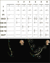
Figure 1
Figure 1
Top, Coronary sinus (CS) electrograms while advancing the CS catheter from subclavian to coronary sinus without using fluoroscopy. Moving from left to right, the panels show first atrial electrograms, then atrial and ventricular electrograms on the distal bipole as the catheter is advanced to the CS os, then atrial and ventricular electrograms on all bipoles confirming CS placement. The electrograms correspond to the positions of the circled numbers below. Bottom, CS catheter displayed on the mapping system to facilitate its placement. Right and left anterior oblique projections of the virtual catheter were displayed. The system reference was used. The bright yellow catheter (No. 6) shows the final CS position and the other shadows record the path of the catheter as it is advanced through the right atrium. The combination of these views and atrial and ventricular electrograms enabled successful CS cannulation in all patients.
Figure 2
Figure 2
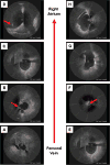
A through D, Serial images as the ICE catheter (within a deflectable sheath) is advanced from the femoral vein to the right atrium. A, Right common iliac vein. B, Junction of the right and left common iliac veins forming the inferior vena cava (IVC). The guide wire from the right side can be seen joining the IVC (red arrow). C, IVC just below the right atrium. D, Right atrium with the ICE catheter located adjacent to the thin-walled fossa ovalis. Often a small amount of curve must be added to the deflectable catheter to bring the ICE catheter this close to the fossa. The bright acoustic artifact of the coronary sinus catheter traversing the right atrium is seen. E through H, Images during passage of the ICE catheter to the right atrium; however, in F, the dramatic reduction in caliber of the vein (arrow) together with palpable resistance illustrates the inadvertent cannulation of a small side branch. By slowly withdrawing the catheter, gently applying a curve to the deflectable sheath, and then rotating the sheath back into the large-caliber lumen of the IVC, the catheter can then be easily advanced to the right atrium.
Figure 3
Figure 3
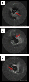
Serial images of the ICE catheter as it is advanced from the right atrium to the innominate vein to prepare the sheath for transseptal puncture. A, Superior vena cava with the coronary sinus (CS) catheter (arrow). B, Inadvertent cannulation of the right innominate vein with the CS catheter (arrow) in the lumen of the left innominate vein. C, ICE catheter is withdrawn and rotated toward the CS catheter (arrow) to follow it into the lumen of the left innominate vein. Subsequently the guide wire, dilator, and tip of the left femoral sheath, all with a characteristic acoustic patterns, can be advanced to this location ready for the transseptal pullback.
Figure 4
Figure 4
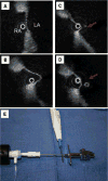
ICE imaging was used to guide the transseptal puncture from right atrium (RA) to LA. The thin membrane of the fossa ovalis (A) and Brockenborough needle tenting the fossa (B) is used to correctly position the needle tip before the puncture. The needle was then advanced as the operator was watching the pressure waveform, feeling for the characteristic “pop,” and looking to see the needle cross into the left atrium on ICE (C, arrow). The dilator and sheath were advanced over the needle until the sheath could be seen in the left atrium (D, arrow). Electrocautery (“cutting,” not “coagulation”) (10 W, <2 seconds) was used to facilitate the transseptal puncture if gentle pressure alone was not successful. The Bovi pen was applied to the back of the Brockenborough needle (E) and power turned on as the needle was advanced out of the tip of the dilator.
Figure 5
Figure 5
Navigation of the ablation catheter in the left atrium was performed using electroanatomic mapping and ICE. A and B, 3D MRI, which has been registered to fiducial points at the ostia of the pulmonary veins using ICE guidance. A, PA view of the left atrium with the green tip of the ablation catheter at the lower posterior aspect of a large left common pulmonary vein. The same position of the ablation catheter is shown in an AP projection of the left atrium in B and on ICE in C. Ablation lesions were marked on the 3D model using red dots. B, The left atrial appendage has been cut away to allow easier viewing of the pulmonary vein behind it. C, The ICE catheter is represented by the white circle in the center of the image. This is located in the center of the lumen of the left common vein. The ablation catheter is the bright artifact in contact with the vein wall directly below the ICE catheter. The esophagus (E) can be seen with its characteristic bright lumen. D, The right superior pulmonary vein with the characteristic pattern of the pulmonary artery (PA) above it and the superior vena cava (SVC), with the coronary sinus catheter (arrow), anterior to this vein. This was a common fiducial point used for registration of the right side of the left atrium.

Similar articles

Cited by

References

    1. McWilliams MJ, Tchou P. The use of a standard radiofrequency energy delivery system to facilitate transseptal puncture. J Cardiovasc Electrophysiol. 2009;20:238–240. - PubMed
    1. Mangrum JM, Mounsey JP, Kok LC, DiMarco JP, Haines DE. Intracardiac echocardiography-guided, anatomically based radiofrequency ablation of focal atrial fibrillation originating from pulmonary veins. J Am Coll Cardiol. 2002;39:1964–1972. - PubMed
    1. Helms A, West JJ, Patel A, Mounsey JP, DiMarco JP, Mangrum JM, Ferguson JD. Real-time rotational ICE imaging of the relationship of the ablation catheter tip and the esophagus during atrial fibrillation ablation. J Cardiovasc Electrophysiol. 2009;20:130–137. - PubMed
    1. Lickfett L, Mahesh M, Vasamreddy C, Bradley D, Jayam V, Eldadah Z, Dickfeld T, Kearney D, Dalal D, Luderitz B, Berger R, Calkins H. Radiation exposure during catheter ablation of atrial fibrillation. Circulation. 2004;110:3003–3010. - PubMed
    1. Limacher MC, Douglas PS, Germano G, Laskey WK, Lindsay BD, McKetty MH, Moore ME, Park JK, Prigent FM, Walsh MN. ACC expert consensus document: radiation safety in the practice of cardiology: American College of Cardiology. J Am Coll Cardiol. 1998;31:892–913. - PubMed