. 2010 Mar;59(3):694-701.
doi: 10.2337/db08-1524. Epub 2009 Dec 15.

Increased vitreous shedding of microparticles in proliferative diabetic retinopathy stimulates endothelial proliferation

Affiliations

Increased vitreous shedding of microparticles in proliferative diabetic retinopathy stimulates endothelial proliferation

Sadri Chahed et al. Diabetes. 2010 Mar.

Abstract

Objective: Diabetic retinopathy is associated with progressive retinal capillary activation and proliferation, leading to vision impairment and blindness. Microparticles are submicron membrane vesicles with biological activities, released following cell activation or apoptosis. We tested the hypothesis that proangiogenic microparticles accumulate in vitreous fluid in diabetic retinopathy.

Research design and methods: Levels and cellular origin of vitreous and plasma microparticles from control (n = 26) and diabetic (n = 104) patients were analyzed by flow cytometry, and their proangiogenic activity was assessed by in vitro thymidine incorporation and neovessel formation in subcutaneous Matrigel plugs in mice.

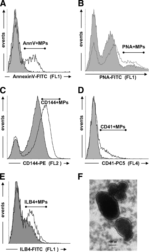
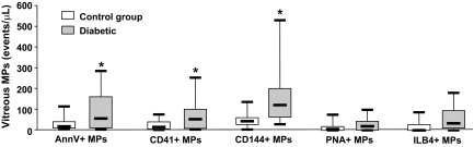
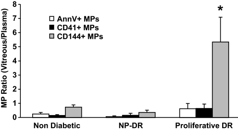
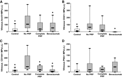
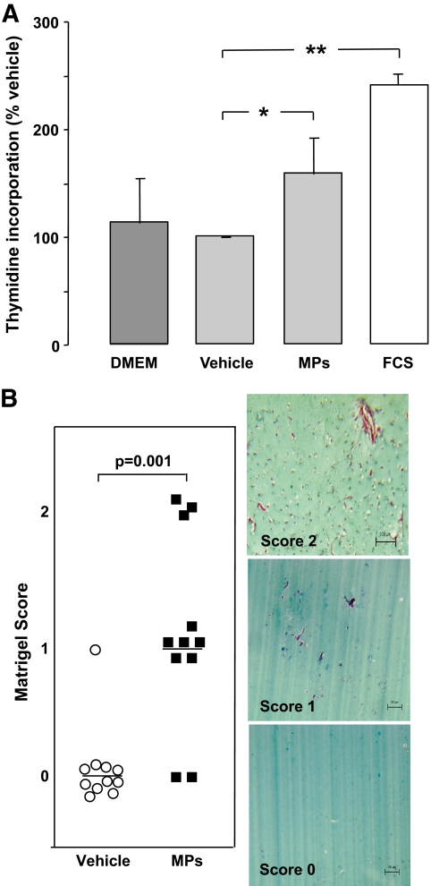
Results: Microparticles of endothelial, platelet, photoreceptor, and microglial origin were identified in vitreous samples. Levels of photoreceptor and microglial microparticles were undetectable in plasmas but were comparable in diabetic and control vitreous samples. Vitreous platelet and endothelial microparticles levels were increased in diabetic patients and decreased following panretinal laser photocoagulation or intravitreal antivascular endothelial growth factor injection in proliferative diabetic retinopathy (PDR). The ratio of vitreous to plasma microparticle levels was calculated to estimate local formation versus potential plasma leakage. In PDR, the endothelial microparticles ratio--but not that for platelet--was greater than 1.0, indicating local formation of endothelial microparticles from retinal vessels and permeation of platelet microparticles from plasma. Isolated vitreous microparticles stimulated by 1.6-fold endothelial proliferation and increased new vessel formation in mice.

Conclusions: The present study demonstrates that vitreous fluid contains shed membrane microparticles of endothelial, platelet, and retinal origin. Vitreous microparticles levels are increased in patients with diabetic retinopathy, where they could contribute to disease progression.


Figures

FIG. 1.
FIG. 1.
Representative traces of flow cytometry analysis of Annexin V+, platelet, endothelial photoreceptor, and microglial microparticles in human vitreous samples. A–E: Size-selected events (0.1–1.0 μm) are plotted as a function of their fluorescence for specific Annexin V–FITC binding (FL1) (A), lectin PNA-FITC labeling (B), anti-human CD144-phycoethrin (PE) labeling (C), anti-human CD41-PC5 labeling (D), or lectin ILB4-FITC labeling (E). Gray-shaded areas represent unspecific labeling determined either in the absence of calcium (A), fluorescent isotypic antibodies (C and D), or d-Galactose (B and E). F: Ultrastructural analyses of vitreous microparticles. Transmission electron microscopic picture of vitreous section showing clustering vitreous vesicles with negative staining. Following samples embedding in Epon 812, thin sections were layed on a carbon-coated grid, stained with 7.6% uranyl acetate and 0.4% lead citrate, and then examined by electron microscopy (Phillips, Eindhoven, the Netherlands; Tecnai, Fei, U.S.). Scale bar: 0.1 μm. FL1, green fluorescence; FL2, orange fluorescence; FL4, infrared fluorescence.
FIG. 2.
FIG. 2.
Vitreous levels of Annexin V+ (AnnV+), platelet (CD41+), endothelial (CD144+), photoreceptor (PNA+) and microglial (ILB4+) microparticles (MPs) in diabetic and control patients. Data are expressed as microparticles/μl and given as median (horizontal bar), 25th and 75th percentile (boxes), and 10th and 90th percentile (error bar). *Significantly different from control patients.
FIG. 3.
FIG. 3.
Ratio of vitreous to plasma levels of Annexin V+ (AnnV+), platelet (CD41+), and endothelial (CD144+) microparticles (MPs) in control and diabetic patients with either PDR or NPDR. *Ratio significantly greater than unity.
FIG. 4.
FIG. 4.
Vitreous levels of Annexin V+ (AnnV+) (A), platelet (CD41+) (B), endothelial (CD144+) (C), and retinal (PNA+) (D) microparticles in control subjects (open bars) and diabetic patients with PDR (gray bars) who underwent either no or incomplete laser panphotocoagulation (PRP), complete PRP, or intravitreal injection of Bevacizumab prior to vitrectomy. *Significantly different when comparing patients with PDR with those with no or incomplete PRP.
FIG. 5.
FIG. 5.
A: HUVECs proliferation assay (n = 4). 3H-thymidine incorporation with the vehicle (vitreous supernatant) was used as the baseline (100%). Levels of 3H-thymidine incorporation are represented with endothelium cell basal medium (Dulbecco's modified Eagle's medium [DMEM]), FCS (2%), and vitreous microparticles (MPs). Vitreous microparticles (estimated as 65 CD144+ microparticles/μl for the endothelial subpopulation) increased 3H-thymidine incorporation by 1.6-fold compared with vehicle (vitreous supernatant without CD144+ microparticles) (*P = 0.029). FCS (10%) increased it by 2.4-fold (**P = 0.003). B: Matrigel scoring and angiogenesis assay. Serial sections of Matrigel were quantified by two independent pathologists. The presence of capillary structures with massive cells invasion was noticed (2). In some plugs, angiogenesis response was limited to cell invasion (1). Low concentration of vitreous microparticles (10 EMP/μl; 5,000 EMPs added to 500 μl Matrigel) induced endothelial cell migration (score 1; n = 6) and new vessel formation (score 2; n = 3) compared with vitreous supernatant (score 0; n = 12) (P = 0.001). (A high-quality digital representation of this figure is available in the online issue.)

References

    1. Frank RN: Diabetic retinopathy. N Engl J Med 2004; 350: 48– 58 - PubMed
    1. Kern TS, Barber AJ: Retinal ganglion cells in diabetes. J Physiol 2008; 586: 4401– 4408 - PMC - PubMed
    1. Curtis TM, Gardiner TA, Stitt AW: Microvascular lesions of diabetic retinopathy: clues towards understanding pathogenesis? Eye 2009; 23: 1496– 1508 - PubMed
    1. Zwaal RF, Schroit AJ: Pathophysiologic implications of membrane phospholipid asymmetry in blood cells. Blood 1997; 89: 1121– 1132 - PubMed
    1. Boulanger CM, Scoazec A, Ebrahimian T, Henry P, Mathieu E, Tedgui A, Mallat Z: Circulating microparticles from patients with myocardial infarction cause endothelial dysfunction. Circulation 2001; 104: 2649– 2652 - PubMed

Publication types

MeSH terms