

doi: 10.1126/scisignal.2101pe78.
Cell mechanics and feedback regulation of actomyosin networks
Affiliations
- PMID: 20009102
- PMCID: PMC4163027
- DOI: 10.1126/scisignal.2101pe78
Item in Clipboard
Cell mechanics and feedback regulation of actomyosin networks
Sci Signal.
.
Abstract
Actomyosin contractility is the major force-generating machinery that shapes cells and tissues during morphogenesis. New evidence from Drosophila demonstrates that these forces are spatially organized by a combination of biochemical and mechanical signals that provide dynamic feedback in a complex cellular environment.
Figures
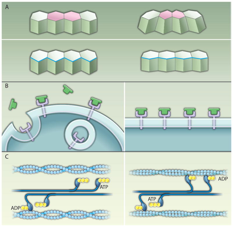
Mechanisms of tension-mediated actomyosin regulation. (A) Actomyosin contraction promotes changes in cell shape during epithelial morphogenesis. During mesoderm invagination and in the amnioserosa during dorsal closure, contraction of an apical actomyosin network (pink) drives apical constriction (top). Leading-edge cells of the lateral epidermis align through the contraction of a multicellular cable (blue) that contributes to dorsal closure (bottom). (B) A model of the plasma membrane as a mechanosensor. In the absence of tension, Fog (green) signaling through its putative receptor (purple) is proposed to be counteracted by endocytosis (left). When the plasma membrane is subjected to external tension, the bending and pinching off of vesicles is inhibited, promoting Fog signaling (9) (right). (C) A model of the myosin II motor as a mechanosensor. For simplicity only one head of each myosin motor is shown. When actin filaments (light blue) are under low tension, myosin II motors in bipolar filaments (dark blue) are rapidly released after adenosine triphosphate (ATP) (yellow) hydrolysis and the power stroke (left). In actin filaments under tension, the ADP bound form of myosin II is stabilized (34-36), leading to the accumulation of myosin II molecules on the actin filament (right).
Similar articles
-
Multiple feedback mechanisms fine-tune Rho signaling to regulate morphogenetic outcomes.J Cell Sci. 2019 Apr 17;132(8):jcs224378. doi: 10.1242/jcs.224378. J Cell Sci. 2019. PMID: 30872456 Free PMC article.
-
Geometry can provide long-range mechanical guidance for embryogenesis.PLoS Comput Biol. 2017 Mar 27;13(3):e1005443. doi: 10.1371/journal.pcbi.1005443. eCollection 2017 Mar. PLoS Comput Biol. 2017. PMID: 28346461 Free PMC article.
-
Planar polarized contractile actomyosin networks in dynamic tissue morphogenesis.Curr Opin Genet Dev. 2017 Aug;45:90-96. doi: 10.1016/j.gde.2017.03.012. Epub 2017 Apr 15. Curr Opin Genet Dev. 2017. PMID: 28419933 Review.
-
The PAR complex regulates pulsed actomyosin contractions during amnioserosa apical constriction in Drosophila.Development. 2010 May;137(10):1645-55. doi: 10.1242/dev.044107. Epub 2010 Apr 14. Development. 2010. PMID: 20392741
-
Forcing cells into shape: the mechanics of actomyosin contractility.Nat Rev Mol Cell Biol. 2015 Aug;16(8):486-98. doi: 10.1038/nrm4012. Epub 2015 Jul 1. Nat Rev Mol Cell Biol. 2015. PMID: 26130009 Free PMC article. Review.
Cited by
-
Willing to Be Involved in Cancer.Genes (Basel). 2016 Jul 18;7(7):37. doi: 10.3390/genes7070037. Genes (Basel). 2016. PMID: 27438856 Free PMC article. Review.
-
Regulation of Cell Behavior by Hydrostatic Pressure.Appl Mech Rev. 2019 Jul;71(4):0408031-4080313. doi: 10.1115/1.4043947. Epub 2019 Jul 23. Appl Mech Rev. 2019. PMID: 31700195 Free PMC article. Review.
-
Role of extracellular matrix and microenvironment in regulation of tumor growth and LAR-mediated invasion in glioblastoma.PLoS One. 2018 Oct 4;13(10):e0204865. doi: 10.1371/journal.pone.0204865. eCollection 2018. PLoS One. 2018. PMID: 30286133 Free PMC article.
-
Why is cytoskeletal contraction required for cardiac fusion before but not after looping begins?Phys Biol. 2015 Jan 30;12(1):016012. doi: 10.1088/1478-3975/12/1/016012. Phys Biol. 2015. PMID: 25635663 Free PMC article.
-
Cell tension and mechanical regulation of cell volume.Mol Biol Cell. 2018 Oct 15;29(21):0. doi: 10.1091/mbc.E18-04-0213. Epub 2018 Aug 16. Mol Biol Cell. 2018. PMID: 30113884 Free PMC article.
References
-
- Dawes-Hoang RE, Parmar KM, Christiansen AE, Phelps CB, Brand AH, Wieschaus EF. Folded gastrulation, cell shape change and the control of myosin localization. Development. 2005;132:4165–4178. - PubMed
-
- Major RJ, Irvine KD. Localization and requirement for Myosin II at the dorsal-ventral compartment boundary of the Drosophila wing. Dev Dyn. 2006;235:3051–3058. - PubMed
-
- Escudero LM, Bischoff M, Freeman M. Myosin II regulates complex cellular arrangement and epithelial architecture in Drosophila. Dev Cell. 2007;13:717–729. - PubMed
Publication types
MeSH terms
Substances
Grants and funding
LinkOut - more resources
Full Text Sources
Molecular Biology Databases
