. 2009;85(10):491-9.
doi: 10.2183/pjab.85.491.

Recombinant alpha-actin for specific fluorescent labeling

Affiliations

Recombinant alpha-actin for specific fluorescent labeling

Atsuko H Iwane et al. Proc Jpn Acad Ser B Phys Biol Sci. 2009.

Abstract

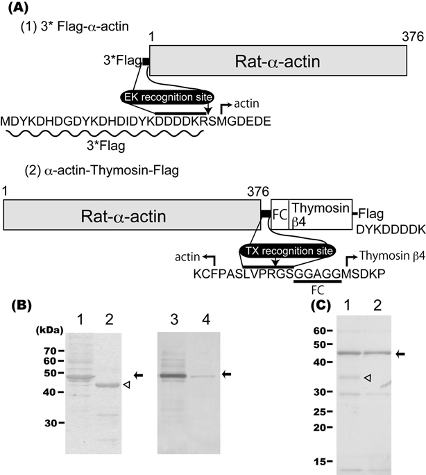
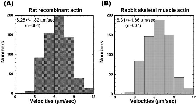
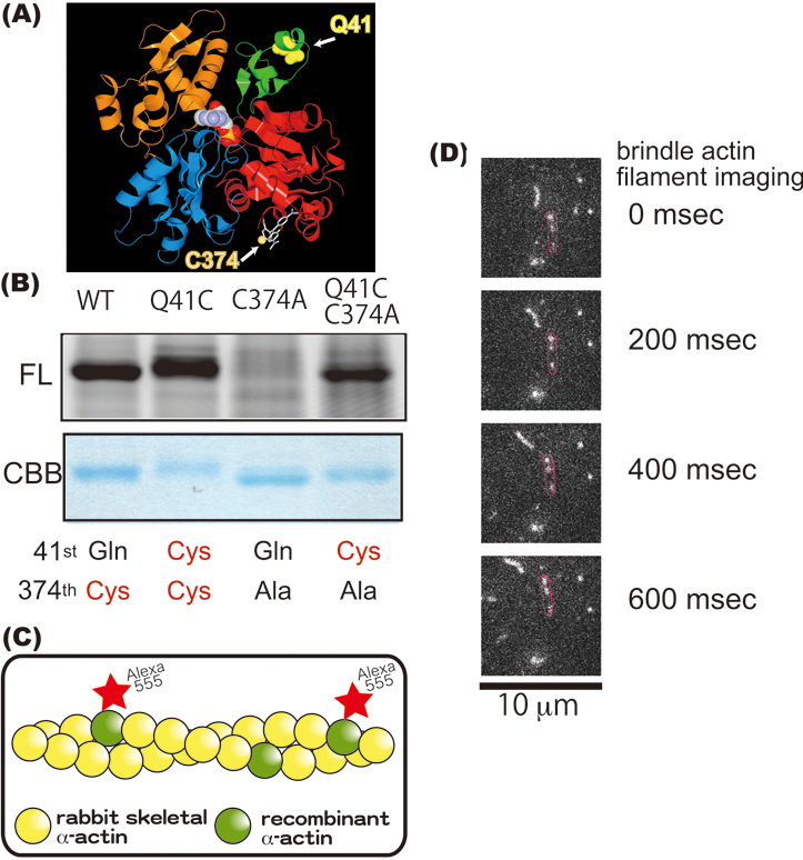
Until recently, actin was thought to act merely as a passive track for its motility partner, myosin, during actomyosin interactions. Yet a recent report having observed dynamical conformational changes in labeled skeletal muscle alpha-actin suggests that actin has a more active role. Because the labeling technique was still immature, however, conclusions regarding the significance of the different conformations are difficult to make. Here, we describe the preparation of fully active alpha-actin obtained from a baculovirus expression system. We developed alpha-actin recombinants, of which subdomains 1 and 2 have specific sites for fluorescent probes. This specific labeling technique offers to significantly expand the information acquired from actin studies.


Figures

Fig. 1.
Fig. 1.
(A) Schematic representation of the two rat α-actin recombinant proteins. FC: glycine rich flexible chain. EK: Enterokinase. TX: Thrombin. Flag: FLAG epitope. Details in text. (B) Purified α-actins with CBB staining before and after enterokinase treatment (lanes 1, 2) and immunoblots using a Flag-tag antibody of the same samples (lanes 3, 4) were loaded on SDS-PAGE (10 percent concentration). Arrows show the position of the 3*Flag-α-actin; arrowhead shows the position of the α-actin after the tag was removed. (C) Purification of α-actin before and after treatment with thrombin protease was checked using SDS-PAGE (12.5 percent concentration) and CBB staining (lanes 1, 2). Arrow and arrowhead point to α-actin and biotinylated thrombin, respectively. For both B and C, approximately 3 μg were loaded onto each lane.
Fig. 2
Fig. 2
(A, B) Histograms of actin filament sliding velocities ((A) rat recombinant α-actin; (B) rabbit skeletal muscle actin) propelled by rabbit HMM directly bound to a nitrocellulose-coated surface. Each histogram shows sliding velocities for >100 individual actin filaments. Assay medium: 20 mM HEPES-KOH (pH 7.8), 25 mM KCl, 5 mM MgCl2, 1 mM ATP and an oxygen scavenger system. Temperature 25 °C.
Fig. 3
Fig. 3
(A) Ribbon representation of the crystal structure of monomeric rabbit α-actin in the ADP state (PDB ID: 1J6z). Four α-actin subdomains are shown (red, subdomain1; green, subdomain2; blue, subdomain3; orange, subdomain4). Residue 41 is seen as a yellow ball while residue 374 is seen as an orange ball via rhodamine. In F-actin, subdomains 1 and 3 point toward the barbed end of the actin filament while subdomains 2 and 4 point toward the pointed end. (B) Analysis of the main band from the SDS-PAGE in Fig. 1. Approximately 2 μg of protein were loaded onto each lane. Upper half gels are fluorescent images of Alexa Fluor 555 C2 maleimide α-actin recombinants; lower half are images stained with CBB. WT is actually our α-actin recombinant. Q41C, C374A and Q41CC374A are α-actin mutants. (C) Schematic drawing of a brindle actin filament. Alexa Fluor 555-labeled recombinant α-actin and rabbit skeletal muscle actin formed a co-filament. (D) Sliding movement of actin co-filaments every 200 miliseconds. The red circles enclose a brindle actin filament.

Similar articles

Cited by

References

    1. Mounier N. and Sparrow J.C. (1997) Structural comparisons of muscle and nonmuscle actins give insights into the evolution of their functional difference. J. Mol. Evo. 44, 89–97 - PubMed
    1. Sellers J.R. (1999) Myosins Second Edition. Oxford University Press, Oxford
    1. Oda T., Iwasa M., Aihara T., Maeda Y. and Narita A. (2009) The nature of the globular- to fibrous-actin transition. Nature 457, 441–445 - PubMed
    1. Kozuka J., Yokota H., Arai Y., Ishii Y. and Yanagida T. (2006) Dynamic polymorphism of single actin molecules in the actin filament. Nat. Chem. Biol. 2, 83–86 - PubMed
    1. Sutoh K., Ando M. and Toyoshima Y.Y. (1991) Site-directed mutations of Dictyostelium actin: disruption of a negative charge cluster at the N terminus. Proc. Natl. Acad. Sci. USA 88, 7711–7714 - PMC - PubMed

Publication types