Review
. 2009 Apr;20(2):79-87.
doi: 10.1097/RMR.0b013e3181c4737e.

Abdominal magnetic resonance elastography

Affiliations
Review

Abdominal magnetic resonance elastography

Meng Yin et al. Top Magn Reson Imaging. 2009 Apr.

Abstract

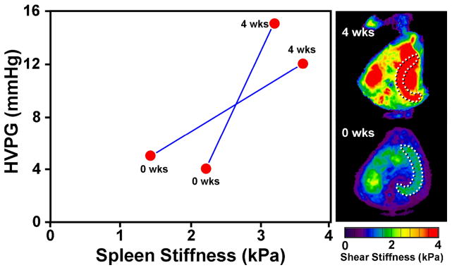
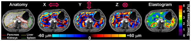
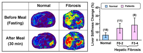
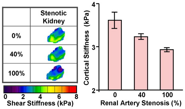
Magnetic resonance elastography (MRE) is a magnetic resonance imaging-based technique for quantitatively assessing the mechanical properties of tissues based on the propagation of shear waves. Multiple studies have described many potential applications of MRE, from characterizing tumors to detecting diffuse disease processes. Studies have shown that MRE can be successfully implemented to assess abdominal organs. The first clinical application of MRE to be well documented is the detection and characterization of hepatic fibrosis, which systematically increases the stiffness of liver tissue. In this diagnostic role, it offers a safer, less expensive, and potentially more accurate alternative to invasive liver biopsy. Emerging results suggest that measurements of liver and spleen stiffness may provide an indirect way to assess portal hypertension. Preliminary studies have demonstrated that it is possible to use MRE to evaluate the mechanical properties of other abdominal structures, such as the pancreas and kidneys. Steady technical progress in developing practical protocols for applying MRE in the abdomen and the pelvis provides opportunities to explore many other potential applications of this emerging technology.


Figures

Figure 1
Figure 1
In an ARPKD mouse model resulting in liver fibrosis, a significant linear correlation was observed between the liver stiffness and the histological analysis of fibrosis extent (p-value < 0.0001). No significant correlation was found between liver stiffness and the fat-to-water ratio in any age group for either the normal or diseased mice. Adapted from reference .
Figure 2
Figure 2
Left: Schematic illustrations of the hepatic MRE setup (with passive driver position indicated) and shear wave generation mechanism; Right: A significant correlation was found between 2-D liver MRE measurements and 3-D liver MRE measurements with full wave information (p < 0.001). Adapted from reference .
Figure 3
Figure 3
Example wave images and shear stiffness maps (elastograms) in liver patients with four different biopsy-proven hepatic fibrosis stages.
Figure 4
Figure 4
Left: The top row shows the results of an MRE exam of a 39-year-old man with a hepatic adenoma. The shear stiffness was measured to be 3.1 kPa in the benign tumor and 2.4 kPa in the surrounding normal liver tissue. The bottom row shows an exam of a 44-year-old man with a cholangiocarcinoma. The stiffness was measured to be 19 kPa in the malignant tumor and 5.4 kPa in the surrounding fibrotic liver tissue. Right: The graph shows a box plot of the shear stiffness of normal liver tissue and various types of hepatic tumors. A cutoff value of 5 kPa separates the malignant tumors from benign tumors and normal liver tissue. Adapted from reference .
Figure 5
Figure 5
Cholestatic liver disease and portal hypertension was established in 20-kg adult mongrel dogs by common bile duct ligation (CBDL). At surgery, two subcutaneous vascular access ports (VAPs) were placed with their catheter tips in the portal vein and right hepatic vein. MRE and measurement of hepatic venous pressure gradient (HVPG) were obtained before and 4 weeks after surgery. In the maximum intensity projection (MIP) elastograms shown on the right, the dotted lines illustrate locations of the canine spleen tissues. The mean spleen stiffness increased substantially (from 1.8 kPa to 3.4 kPa) with the severely elevated mean HVPG (from 4.5 mmHg to 13.5 mmHg).
Figure 6
Figure 6
One slice from a 3-D/3-axis abdominal MRE exam of a healthy volunteer showing excellent depictions of the liver, spleen, pancreas, and kidneys.
Figure 7
Figure 7
Left: Examples of hepatic MRE exams performed pre- and postprandially in a normal volunteer and in a patient with cirrhotic liver. The liver stiffness in the patient with the cirrhotic liver was increased markedly in the postprandial exam. Right: A summary of the percent change in liver stiffness for healthy volunteers and patients with various stages of fibrosis. Results in 18 normal subjects demonstrated no significant change in postprandial hepatic stiffness compared with the fasting state. However, results obtained in 19 patients with hepatic fibrosis demonstrated a statistically significant increase in postprandial liver stiffness (p<0.01).
Figure 8
Figure 8
An acute RAS model was established in a 45-kg adult pig by isolating the renal artery to insert a vascular occluder and an embedded Doppler flow probe. The RBF was reduced from the baseline level to 40% and then to total occlusion of 100%. The MRE-assessed renal tissue stiffness measurements demonstrated that the renal tissue stiffness decreases during gradual reductions in RBF in pigs.

References

    1. Lerner RM, Huang SR, Parker KJ. “Sonoelasticity” images derived from ultrasound signals in mechanically vibrated tissues. Ultrasound Med Biol. 1990;16(3):231–239. - PubMed
    1. Sandrin L, Tanter M, Gennisson JL, Catheline S, Fink M. Shear elasticity probe for soft tissues with 1-D transient elastography. IEEE Trans Ultrason Ferroelectr Freq Control. 2002 Apr;49(4):436–446. - PubMed
    1. Muthupillai R, Lomas DJ, Rossman PJ, Greenleaf JF, Manduca A, Ehman RL. Magnetic resonance elastography by direct visualization of propagating acoustic strain waves. Science. 1995 Sep 29;269(5232):1854–1857. - PubMed
    1. Bernstein MA, Ikezaki Y. Comparison of phase-difference and complex-difference processing in phase-contrast MR angiography. J Magn Reson Imaging. 1991;1:725–729. - PubMed
    1. Muthupillai R, Rossman PJ, Lomas DJ, Greenleaf JF, Riederer SJ, Ehman RL. Magnetic resonance imaging of transverse acoustic strain waves. Magn Reson Med. 1996 Aug;36(2):266–274. - PubMed

MeSH terms