

Retinal pigment epithelial changes in chronic Vogt-Koyanagi-Harada disease: fundus autofluorescence and spectral domain-optical coherence tomography findings
- PMID: 20010321
- PMCID: PMC2903055
- DOI: 10.1097/IAE.0b013e3181c5970d
Retinal pigment epithelial changes in chronic Vogt-Koyanagi-Harada disease: fundus autofluorescence and spectral domain-optical coherence tomography findings
Abstract
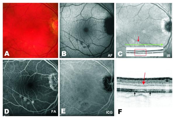
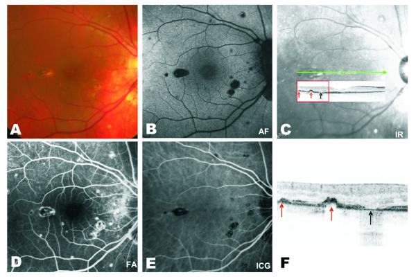
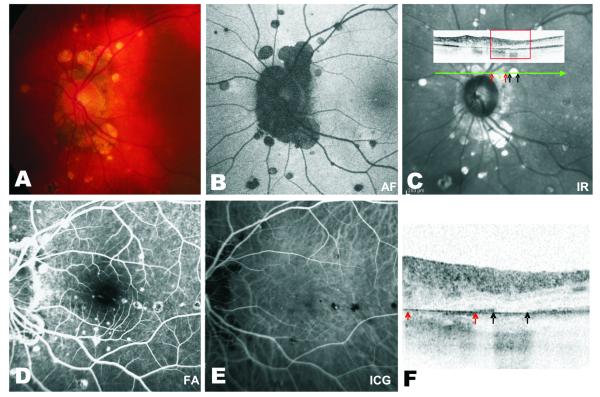
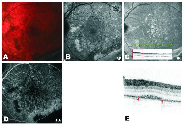
Purpose: The purpose of this study was to determine whether fundus autofluorescence (FAF) and spectral domain-optical coherence tomography (SD-OCT) imaging allow better assessment of retinal pigment epithelium and the outer retina in subjects with chronic Vogt-Koyanagi-Harada disease compared with examination and angiography alone.
Methods: A cross-sectional analysis of a series of seven consecutive patients with chronic Vogt-Koyanagi-Harada disease undergoing FAF and SD-OCT was conducted. Chronic disease was defined as duration of intraocular inflammation >3 months. Color fundus photographs were correlated to FAF and SD-OCT images. The images were later correlated to fluorescein angiography and indocyanine green angiography.
Results: All patients had sunset glow fundus, which resulted in no apparent corresponding abnormality on FAF or SD-OCT. Lesions with decreased autofluorescence signal were observed in 11 eyes (85%), being associated with loss of the retinal pigment epithelium and involvement of the outer retina on SD-OCT. In 5 eyes (38%), some of these lesions were very subtle on clinical examination but easily detected by FAF. Lesions with increased autofluorescence signal were seen in 8 eyes (61.5%), showing variable involvement of the outer retina on SD-OCT and corresponding clinically to areas of retinal pigment epithelium proliferation and cystoid macular edema.
Conclusion: Combined use of FAF and SD-OCT imaging allowed noninvasive delineation of retinal pigment epithelium/outer retina changes in patients with chronic Vogt-Koyanagi-Harada disease, which were consistent with previous histopathologic reports. Some of these changes were not apparent on clinical examination.
Figures
References
-
- Moorthy RS, Inomata H, Rao NA. Vogt-Koyanagi-Harada syndrome. Surv Ophthalmol. 1995;39:265–292. - PubMed
-
- Rao NA. Mechanisms of inflammatory response in sympathetic ophthalmia and VKH syndrome. Eye. 1997;11:213–216. - PubMed
-
- Read RW, Holland GN, Rao NA, et al. Revised diagnostic criteria for Vogt-Koyanagi-Harada disease: report of an international committee on nomenclature. Am J Ophthalmol. 2001;131:647–652. - PubMed
-
- Beniz J, Forster DJ, Lean JS, et al. Variations in clinical features of the Vogt-Koyanagi-Harada syndrome. Retina. 1991;11:275–280. - PubMed
-
- Read RW, Yu F, Accorinti M, et al. Evaluation of the effect on outcomes of the route of administration of corticosteroids in acute Vogt-Koyanagi-Harada disease. Am J Ophthalmol. 2006;142:119–124. - PubMed
Publication types
MeSH terms
Substances
Grants and funding
LinkOut - more resources
Full Text Sources
