. 2010 Feb 3;29(3):606-18.
doi: 10.1038/emboj.2009.369. Epub 2009 Dec 10.

Antagonism of Beclin 1-dependent autophagy by BCL-2 at the endoplasmic reticulum requires NAF-1

Affiliations

Antagonism of Beclin 1-dependent autophagy by BCL-2 at the endoplasmic reticulum requires NAF-1

Natasha C Chang et al. EMBO J. .

Abstract

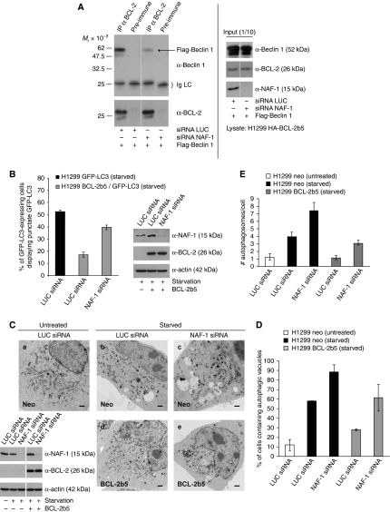
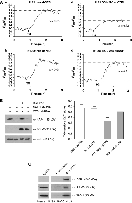
In addition to mitochondria, BCL-2 is located at the endoplasmic reticulum (ER) where it is a constituent of several distinct complexes. Here, we identify the BCL-2-interacting protein at the ER, nutrient-deprivation autophagy factor-1 (NAF-1)-a bitopic integral membrane protein whose defective expression underlies the aetiology of the neurodegenerative disorder Wolfram syndrome 2 (WFS2). NAF-1 contains a two iron-two sulphur coordinating domain within its cytosolic region, which is necessary, but not sufficient for interaction with BCL-2. NAF-1 is displaced from BCL-2 by the ER-restricted BH3-only protein BIK and contributes to regulation of BIK-initiated autophagy, but not BIK-dependent activation of caspases. Similar to BCL-2, NAF-1 is found in association with the inositol 1,4,5-triphosphate receptor and is required for BCL-2-mediated depression of ER Ca(2+) stores. During nutrient deprivation as a physiological stimulus of autophagy, BCL-2 is known to function through inhibition of the autophagy effector and tumour suppressor Beclin 1. NAF-1 is required in this pathway for BCL-2 at the ER to functionally antagonize Beclin 1-dependent autophagy. Thus, NAF-1 is a BCL-2-associated co-factor that targets BCL-2 for antagonism of the autophagy pathway at the ER.


Conflict of interest statement

The authors declare that they have no conflict of interest.

Figures

Figure 1
Figure 1
Identification of a novel BCL-2-interacting partner, NAF-1, at the ER. (A) BH3-only BIK displaces a subset of BCL-2 complexes at the ER. Purified LM from H1299 HA-BCL-2b5 cells either mock infected or infected with Ad-BIKb5 was subjected to cross-linking with BMH and visualized by immunoblot. Asterisks (*) denote BCL-2 cross-linked products, which are displaced by BIK. Bold arrow denotes region containing cross-linked NAF-1/BCL-2. (B) Amino-acid sequence of NAF-1. Peptide sequences identified by mass spectrometry are indicated by a double underline. The TM domain is indicated by a bold single underline. Cys residues in the cytosolic domain are denoted with asterisks (*). The CDGSH domain is highlighted in grey, and the predicted 2Fe-2S coordinating Cys-3-His-1 aa are marked in bold. A downwards arrow head (∨) represents the site in which a Flag epitope was inserted. A double-lined arrow (⇓) indicates the Glu residue that when mutated (E37Q) causes WFS2. Predicted phosphorylation sites found using the PhosphoMotif Finder from the Human Protein Reference Database (HPRD) are indicated in bold within a box; β-adrenergic receptor kinase substrate motif (aa 25–29), PKA and PKC kinase substrate motifs (aa 32–34 and 67–69), and casein kinase II substrate motif (aa 34–37). (C) Deduced membrane topology of NAF-1. Grey rectangle indicates CDGSH domain. (D) Endogenous NAF-1 cross-links with BCL-2 at the ER. LM purified from H1299 neo and HA-BCL-2b5 cells were subjected to cross-linking by BMH and immunoprecipitation with anti-NAF-1 antibody. Precipitates were analysed by immunoblot with anti-BCL-2. (E) NAF-1 is displaced from BCL-2 by BH3-only BIK. H1299 HA-BCL-2b5 cells were either mock infected or infected with Ad-BIK. Purified LM was treated as in (D).
Figure 2
Figure 2
The NAF-1/BCL-2 interaction. (A) Co-immunoprecipitation of endogenous NAF-1 and BCL-2. Lysates from SK-Mel5 cells were collected and immunoprecipitation was performed with anti-NAF-1 antibody. The precipitate was subjected to immunoblotting with anti-BCL-2 and anti-NAF-1. (B) H1299 cells were fixed and double stained with anti-NAF-1 and anti-Calnexin or anti-Cytochrome c antibodies. Scale bar represents 10 μm. (C) Co-immunoprecipitation of NAF-1-Flag and HA-BCL-2b5. Lysates from H1299 HA-BCL-2b5 cells infected with Ad-NAF-1-Flag were collected and treated as in (A). (D) Mutations in the CDGSH iron-binding domain of NAF-1 interfere with NAF-1 binding to BCL-2. H1299 HA-BCL-2b5 cells were infected with either Ad-rtTA, Ad-NAF-1-Flag, or Ad-NAF-1-mut-Flag (C99S C101S C110S H114Q). Lysates were treated as in (A). Densitometric analysis was performed using Scion Image software to quantify expression and co-precipitated levels of NAF-1-Flag and NAF-1-mut-Flag. Graph depicts the ratio of co-precipitated protein to expression level. (E) A functional CDGSH iron-binding domain is necessary, but not sufficient for the interaction between the cytosolic domains of NAF-1 and BCL-2. HA-BCL-2 ΔTM was in vitro translated in rabbit reticulocyte lysate and equivalent aliquots were added to each GST pull-down reaction. GST-fusion proteins used were GST alone, GST-NAF1-C, GST-NAF1-C-mut (C99S C101S C110S H114Q), and GST-MitoNEET-C. The proteins were detected using anti-HA and anti-GST.
Figure 3
Figure 3
NAF-1 does not affect BIK-initiated apoptosis, but influences BIK-induced autophagy in the absence of caspase activation. (A) H1299 neo and HA-BCL-2b5 cells treated with control (CTRL) or NAF-1 shRNA were either mock infected or infected with Ad-BIK in the absence or presence of 50 μM zVAD-fmk. Cell lysates were analysed by immunoblot. (B) Caspase activity was measured using the fluorescent substrate DEVD-AMC. The results represent the average±s.d. of three independent experiments. (C) Prolonged BIK expression and caspase inhibition induces autophagy, which is enhanced by knockdown of NAF-1. H1299 neo and HA-BCL-2b5 cells treated with CTRL or NAF-1 shRNA were infected with Ad-BIK in the presence of zVAD-fmk for the indicated periods of time. Cell lysates were analysed by immunoblot. Levels of LC3 II were normalized to actin levels by densitometry analysis. Graph depicts normalized LC3 II levels of each lane.
Figure 4
Figure 4
NAF-1 knockdown results in increased incidence and intensity of starvation-induced Beclin 1-dependent autophagy. (A) Effect of NAF-1 knockdown on starvation-induced autophagy. H1299 cells infected with CTRL or NAF-1 shRNA were starved for 4 h in EBSS with DMSO (vehicle) or Baf A1 (100 nM). Cell lysates were analysed by immunoblot. (B) Representative images of GFP-LC3 staining in H1299 GFP-LC3 cells transfected with LUC or NAF-1 siRNA, untreated and starved. Scale bar represents 10 μm. (C) Quantification of autophagy is expressed as the percentage of GFP-LC3-expressing cells displaying punctate GFP-LC3. A minimum of 100 cells per sample were counted; results represent the average±s.d. of three independent experiments. Cell lysates were analysed by immunoblot.
Figure 5
Figure 5
BCL-2 inhibition of Beclin 1-dependent autophagy requires NAF-1. (A) NAF-1 contributes to the interaction between BCL-2 and Beclin 1. H1299 HA-BCL-2b5 cells were transfected with Flag-Beclin 1 and either LUC or NAF-1 siRNA. Cells were lysed and subjected to immunoprecipitation with anti-BCL-2 antibody. Precipitates were subjected to analysis by immunoblot using anti-Beclin 1 and anti-BCL-2. All lanes are derived from the same gel and of the same exposure. Thin white lines indicate where lanes have been removed. (B) Loss of NAF-1 prevents BCL-2b5 from antagonizing starvation-induced autophagy. H1299 GFP-LC3 and BCL-2b5/GFP-LC3 cells were transfected with either LUC or NAF-1 siRNA and starved for 4 h. Cells were analysed as in Figure 4C. (C) Representative electron micrographs of H1299 neo and HA-BCL-2b5 cells treated with either LUC or NAF-1 siRNA, with or without subsequent starvation. Scale bar represents 10 μm. Cell lysates were analysed by immunoblot. All lanes are derived from the same gel and of the same exposure. Thin white line indicates where lanes have been removed. Autophagy levels observed by electron microscopy (C) were quantified and expressed as either the percentage of cells containing autophagic vacuoles (D) or the number of autophagosomes per cell (E). A minimum of 100 cells per sample were counted; results represent the average±s.e.m.
Figure 6
Figure 6
NAF-1 knockdown reverses BCL-2 lowering of ER Ca2+ stores. (A) H1299 neo and HA-BCL-2b5 cells treated with either CTRL or NAF-1 shRNA were loaded with Fura-2AM, and ER Ca2+ stores were measured as the difference in cytoplasmic Ca2+ concentration before and after addition of TG (2 μM). Shown are representative traces of Fura-2AM fluorescence measured at 340/380 nm excitation wavelength ratio at 510 nm wavelength emission. Arrow indicates time at which TG was added, delta values indicate TG-sensitive ER Ca2+ stores. (B) Differences in ER Ca2+ stores are shown as the average±s.e.m. of three independent experiments as described in (A). (C) Co-immunoprecipitation of endogenous NAF-1 and endogenous IP3 receptor type I. Lysate from H1299 HA-BCL-2b5 cells was collected and immunoprecipitation was performed with anti-IP3R1 antibody. The resulting precipitate was analysed by immunoblot.

Comment in

References

    1. Amr S, Heisey C, Zhang M, Xia XJ, Shows KH, Ajlouni K, Pandya A, Satin LS, El-Shanti H, Shiang R (2007) A homozygous mutation in a novel zinc-finger protein, ERIS, is responsible for Wolfram syndrome 2. Am J Hum Genet 81: 673–683 - PMC - PubMed
    1. Axe EL, Walker SA, Manifava M, Chandra P, Roderick HL, Habermann A, Griffiths G, Ktistakis NT (2008) Autophagosome formation from membrane compartments enriched in phosphatidylinositol 3-phosphate and dynamically connected to the endoplasmic reticulum. J Cell Biol 182: 685–701 - PMC - PubMed
    1. Boyd JM, Gallo GJ, Elangovan B, Houghton AB, Malstrom S, Avery BJ, Ebb RG, Subramanian T, Chittenden T, Lutz RJ, Chinnadurai G (1995) Bik, a novel death-inducing protein shares a distinct sequence motif with Bcl-2 family proteins and interacts with viral and cellular survival-promoting proteins. Oncogene 11: 1921–1928 - PubMed
    1. Brady NR, Hamacher-Brady A, Yuan H, Gottlieb RA (2007) The autophagic response to nutrient deprivation in the hl-1 cardiac myocyte is modulated by Bcl-2 and sarco/endoplasmic reticulum calcium stores. FEBS J 274: 3184–3197 - PubMed
    1. Breckenridge DG, Nguyen M, Kuppig S, Reth M, Shore GC (2002) The procaspase-8 isoform, procaspase-8L, recruited to the BAP31 complex at the endoplasmic reticulum. Proc Natl Acad Sci USA 99: 4331–4336 - PMC - PubMed

Publication types

MeSH terms