Review
. 2010 Jan;10(1):11-23.
doi: 10.1038/nri2674. Epub 2009 Dec 11.

The immune response during acute HIV-1 infection: clues for vaccine development

Affiliations
Review

The immune response during acute HIV-1 infection: clues for vaccine development

Andrew J McMichael et al. Nat Rev Immunol. 2010 Jan.

Abstract

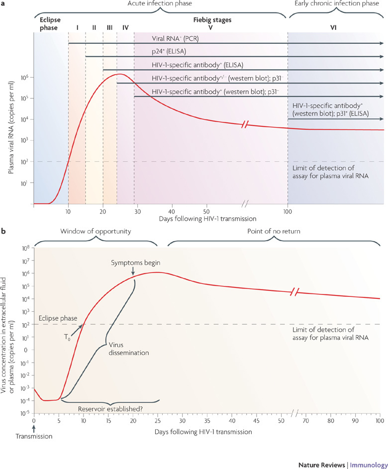
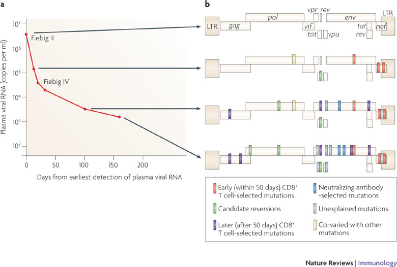
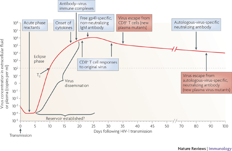
The early immune response to HIV-1 infection is likely to be an important factor in determining the clinical course of disease. Recent data indicate that the HIV-1 quasispecies that arise following a mucosal infection are usually derived from a single transmitted virus. Moreover, the finding that the first effective immune responses drive the selection of virus escape mutations provides insight into the earliest immune responses against the transmitted virus and their contributions to the control of acute viraemia. Strong innate and adaptive immune responses occur subsequently but they are too late to eliminate the infection. In this Review, we discuss recent studies on the kinetics and quality of early immune responses to HIV-1 and their implications for developing a successful preventive HIV-1 vaccine.


Conflict of interest statement

The authors declare no competing financial interests.

Figures

Figure 1
Figure 1. Definition of acute HIV-1 infection.
a | Recent analysis of samples from individuals early after infection with HIV-1 has revealed that the first weeks following infection can be divided into clinical stages that are defined by a stepwise gain in positivity for the detection of HIV-1 antigens and HIV-1-specific antibodies in diagnostic assays (in brackets). The time between infection and the first detection of viral RNA in the plasma is referred to as the eclipse phase. Plasma virus levels then increase exponentially, peaking at 21–28 days after infection, and this is followed by a slower decrease in plasma viral RNA levels. Patients can be categorized into Fiebig stages I–VI, which are based on a sequential gain in positive HIV-1 clinical diagnostic assays (viral RNA measured by PCR, p24 and p31 viral antigens measured by enzyme-linked immunosorbent assay (ELISA), HIV-1-specific antibody detected by ELISA and HIV-1-specific antibodies detected by western blot). Patients progress from acute infection through to the early chronic stage of infection at the end of Fiebig stage V, approximately 100 days following infection, as the plasma viral load begins to plateau. b | Fundamental events in acute HIV-1 infection. Following HIV-1 infection, the virus first replicates locally in the mucosa and is then transported to draining lymph nodes, where further amplification occurs. This initial phase of infection, until systemic viral dissemination begins, constitutes the eclipse phase. The time when virus is first detected in the blood is referred to as T0; after this there is an exponential increase in plasma viraemia to a peak 21–28 days after infection. By this time, significant depletion of mucosal CD4+ T cells has already occurred. Around the time of peak viraemia, patients may become symptomatic and reservoirs of latent virus are established in cells that have a slower rate of decay than CD4+ T cells. The 'window of opportunity' between transmission and peak viraemia, prior to massive CD4+ T cell destruction and the establishment of viral reservoirs, is the narrow but crucial period in which an HIV-1 vaccine must control viral replication, prevent extensive CD4+ T cell depletion and curb generalized immune activation. Part a is modified, with permission, from Ref. © (2008) National Academy of Sciences, USA. Part b is modified from Ref. .
Figure 2
Figure 2. Early events in acute HIV-1 infection.
a | The timing of the appearance of soluble proteins and apoptotic microparticles in the blood during acute HIV-1 infection. An increase in the level of soluble tumour necrosis factor-related apoptosis-inducing ligand (TRAIL; also known as TNFSF10) in the plasma is the first evidence of infection-induced apoptosis and/or immune activation and it occurs before the peak in viraemia. This increase in TRAIL also coincides with the appearance of high levels of interferon-α (IFNα; see Fig. 3). Soon thereafter increases in the number of apoptotic microparticles are observed, followed by increased levels of soluble TNF receptor 2 (TNFR2; also known as TNFRSF1B) and soluble FAS ligand. A portion of the microparticles express CC-chemokine receptor 5, suggesting that they originate from cellular targets of HIV-1 infection. The appearance of these soluble components in the plasma early during HIV-1 infection probably represents a pathological rather than a protective cascade of events that is a cause of, or is associated with, virus-induced CD4+ T cell death. b | Electron micrograph of apoptotic microparticles and exosomes in the plasma from a patient with acute HIV-1 infection. There are ∼700-fold more microparticles than virions present in acute HIV-1 infection. Part a is based on data from Ref. . Part b is reproduced, with permission, from Ref. © (2008) American Society for Microbiology.
Figure 3
Figure 3. The cytokine storm in acute HIV-1 infection.
The relative kinetics of elevation of acute-phase proteins, cytokines and chemokines in the plasma during acute HIV-1 infection. There are two initial waves of cytokines: interleukin-15 (IL-15) and interferon-α (IFNα), followed by tumour necrosis factor (TNF), IL-18 and IL-10. CXCL10, CXC-chemokine ligand 10. Figure is reproduced, with permission, from Ref. © (2009) American Society for Microbiology.
Figure 4
Figure 4. Early T cell selection of virus escape mutations in acute HIV-1 infection.
The virus escape mutations occurring in a single representative patient during acute HIV-1 infection. The changes in plasma virus load (a) and the emergence of amino acid changes (b) are shown. At the first time point, when the patient was virus positive but seronegative (Fiebig stage II), the founder virus (which was clade B) showed no evidence of immune selection. Thereafter there is an increasing number of selected sites at which the founder virus sequence is completely altered by, usually a cluster of, amino acid changes. Those marked in red were selected early (within 50 days from peak viraemia) by demonstrable CD8+ T cell responses. Those in purple were selected later by CD8+ T cells. Those in light green are single amino acid reversions to the clade B virus consensus sequence. Those in blue were mutations in V1 and V3 of the env gene selected by neutralizing antibodies. Those in grey were selected through undefined means, possibly by T cells, natural killer cells or antibodies. Yellow represents changes that co-varied with another mutation. LTR, long terminal repeat. Figure is based on data from Ref. .
Figure 5
Figure 5. Composite alignment of the earliest innate and adaptive immune responses detected after HIV-1 transmission.
The first systemically detectable immune responses to HIV-1 infection are the increases in levels of acute-phase proteins in the plasma, which are observed when virus replication is still largely restricted to the mucosal tissues and draining lymph nodes (eclipse phase). When virus is first detected in the plasma (T0), broad and dynamic increases in plasma cytokine levels are also observed. Within days, as plasma viraemia is still increasing exponentially, the first antibody–virus immune complexes are detected. Expansion of the earliest HIV-1-specific CD8+ T cell responses also commences prior to peak viraemia, followed by detection of the first free glycoprotein (gp)41-specific but non-neutralizing IgM antibodies. Complete virus escape from the first CD8+ T cell responses can occur rapidly, within 10 days of T cell expansion. By this time, viral reservoirs exist, possibly becoming established within days of infection. The earliest autologous-virus-neutralizing antibodies are detected around day 80 following infection, as viral loads are still declining prior to the onset of the viral set point. Antibody escape virus mutants emerge in the plasma within the following week.

Similar articles

Cited by

References

    1. Pilcher CD, et al. Detection of acute infections during HIV testing in North Carolina. N. Engl. J. Med. 2005;352:1873–1883. doi: 10.1056/NEJMoa042291. - DOI - PubMed
    1. Powers KA, et al. Improved detection of acute HIV-1 infection in sub-Saharan Africa: development of a risk score algorithm. AIDS. 2007;21:2237–2242. doi: 10.1097/QAD.0b013e3282f08b4d. - DOI - PMC - PubMed
    1. Hu Q, et al. Blockade of attachment and fusion receptors inhibits HIV-1 infection of human cervical tissue. J. Exp. Med. 2004;199:1065–1075. doi: 10.1084/jem.20022212. - DOI - PMC - PubMed
    1. Veazey RS, et al. Protection of macaques from vaginal SHIV challenge by vaginally delivered inhibitors of virus-cell fusion. Nature. 2005;438:99–102. doi: 10.1038/nature04055. - DOI - PubMed
    1. Margolis L, Shattock R. Selective transmission of CCR5-utilizing HIV-1: the 'gatekeeper' problem resolved? Nature Rev. Microbiol. 2006;4:312–317. doi: 10.1038/nrmicro1387. - DOI - PubMed

Publication types