. 2010 Jan;12(1):66-73; sup pp 1-9.
doi: 10.1038/ncb2006. Epub 2009 Dec 13.

A bacterial E3 ubiquitin ligase IpaH9.8 targets NEMO/IKKgamma to dampen the host NF-kappaB-mediated inflammatory response

Affiliations

A bacterial E3 ubiquitin ligase IpaH9.8 targets NEMO/IKKgamma to dampen the host NF-kappaB-mediated inflammatory response

Hiroshi Ashida et al. Nat Cell Biol. 2010 Jan.

Abstract

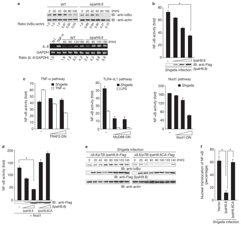
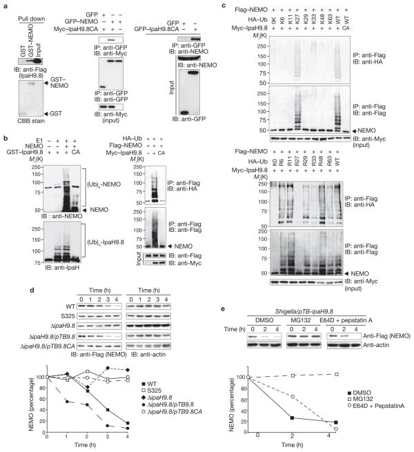
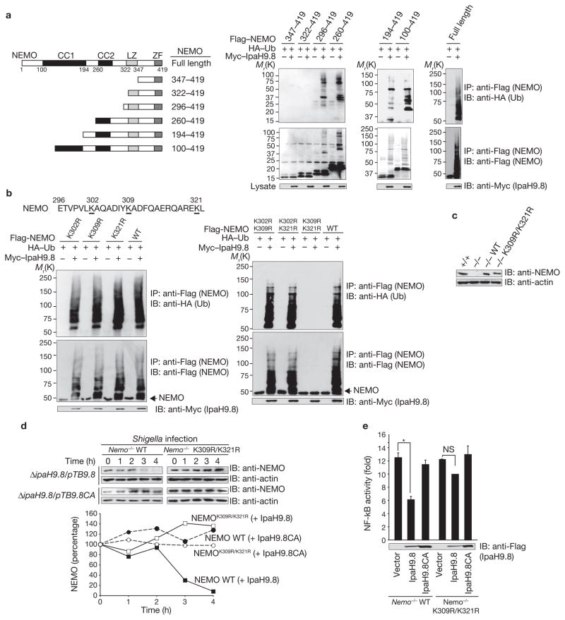
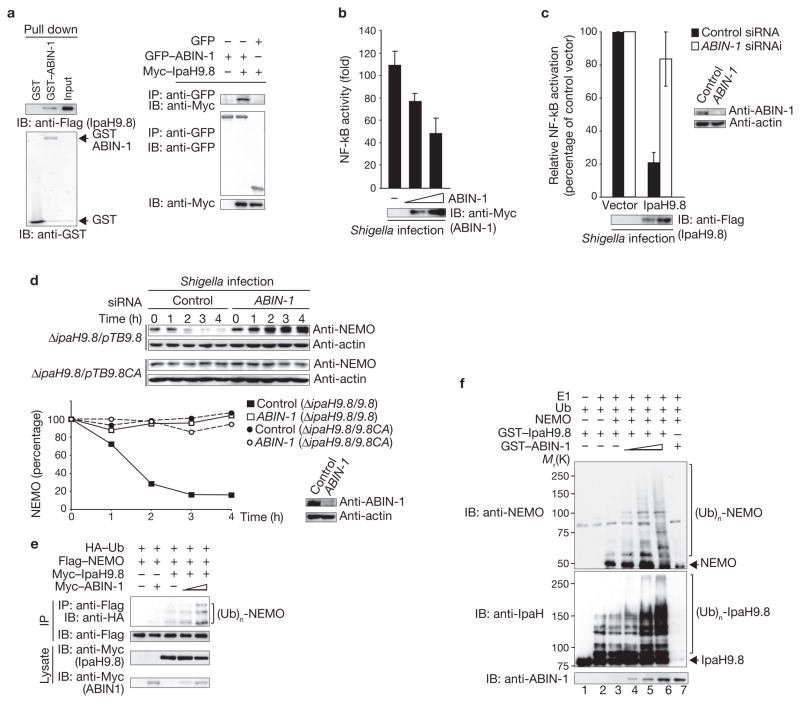
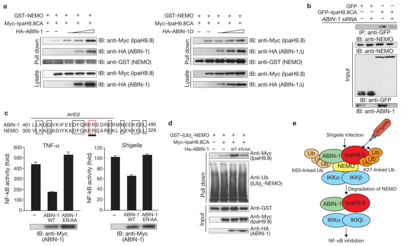
NF-kappaB (nuclear factor kappaB) has a pivotal role in many cellular processes, including the inflammatory and immune responses and, therefore, its activation is tightly regulated by the IKK (IkappaB kinase) complex and by IkappaBalpha degradation. When Shigella bacteria multiply within epithelial cells they release peptidoglycans, which are recognized by Nod1 and stimulate the NF-kappaB pathway, thus leading to a severe inflammatory response. Here, we show that IpaH9.8, a Shigella effector possessing E3 ligase activity, dampens the NF-kappaB-mediated inflammatory response to the bacterial infection in a unique way. IpaH9.8 interacts with NEMO/IKKgamma and ABIN-1, a ubiquitin-binding adaptor protein, promoting ABIN-1-dependent polyubiquitylation of NEMO. Consequently, polyubiquitylated NEMO undergoes proteasome-dependent degradation, which perturbs NF-kappaB activation. As NEMO is essential for NF-kappaB activation, we propose that the polyubiquitylation and degradation of NEMO during Shigella infection is a new bacterial strategy to modulate host inflammatory responses.


Conflict of interest statement

COMPETING FINANCIAL INTERESTS

The authors declare no competing financial interests.

Figures

Figure 1
Figure 1
IpaH9.8 inhibits NF-κB activity. (a) HeLa cells were infected with Shigella wild-type or ΔipaH9.8 mutant bacteria harbouring an afimbrial adhesin (Afa) expression plasmid (multiplicity of infection, MOI = 10). After Shigella invasion, cell lysates were prepared at the indicated time points and subjected to immunoblotting (upper panel) or semi-quantitative reverse transcription-PCR (RT-PCR) analysis (lower panel). The blots were quantified by measuring relative intensity, and values are indicated below the blots. (b) Luciferase assays were performed after Shigella infection (for 3 h) of 293T cells transiently transfected with an NF-κB reporter plasmid (50 ng) and an empty vector or a Flag–IpaH9.8-expressing plasmid (0, 50, 100 or 200 ng). Results are presented as fold relative to the activity of non-infected cells. Data are mean ± s.e.m., n = 4. (c) Luciferase assays of 293T cells transiently transfected with an NF-κB reporter plasmid and an empty vector or increasing amounts of dominant-negative forms of TRAF2 (TRAF2-DN), MyD88 (MyD88-DN) or Nod1 (Nod1-DN). After 24 h, cells were infected with Shigella or treated with TNF-α (10 ng ml−1) or LPS (100 ng ml−1) for 3 h and luciferase activity was measured. Results are presented as fold relative to the activity of non-infected or non-stimulated cells. Data are mean ± s.e.m., n = 3. (d) Luciferase assay of 293T cells transfected with an NF-κB reporter plasmid (50 ng) and empty vector, Flag–IpaH9.8 or Flag–IpaH9.8CA expressing plasmid (50 or 200 ng), plus plasmids expressing Nod1 as an NF-κB activator. Results are presented as fold relative to the activity of non-stimulated cells. Data are mean ± s.e.m., n = 4. (e) HeLa cells were infected with Shigella ΔipaH9.8 harbouring pTB-ipaH9.8–Flag or pTB-ipaH9.8CAFlag (pTB is an IPTG-inducible vector). The cell lysates prepared at the indicated time points were subjected to immunoblotting. (f) The percentage of nuclear translocation of NF-κB (p65) after Shigella infection for 1 h was determined by counting the number of nuclear translocated p65 in at least 300 GFP-, GFP–IpaH9.8- or GFP–IpaH9.8CA-expressing cells using fluorescence microscopy. Data are mean ± s.e.m., n = 3. *P < 0.001. WT, wild-type. IB, immunoblot. N.I, non-infected. For full scans of blots in a, b, d and e, see Supplementary Information, Fig. S7.
Figure 2
Figure 2
IpaH9.8 promotes NEMO ubiquitylation and degradation. (a) IpaH9.8–Flag bound to NEMO, as found in GST pulldown assays (left panel) and immunoprecipitation analysis of 293T cell lysates (center and right panel). (b) IpaH9.8 targets NEMO, and promotes its ubiquitylation. In vitro ubiquitylation assay with NEMO and a mixture of E1, UbcH5b, ATP and ubiquitin in the presence or absence of GST–IpaH9.8 or GST–IpaH9.8CA (left panel). Cells expressing Flag–NEMO, HA–Ub and Myc6–IpaH9.8 or Myc6–IpaH9.8CA were immunoprecipitated using an anti-Flag antibody and analysed for ubiquitylation by immunoblotting using anti-HA and anti-Flag antibodies. Whole cell lysates were immunoblotted with anti-Flag and anti-Myc antibodies (right panel). (c) Cells expressing Flag–NEMO, Myc6-IpaH9.8 and each of the HA-ubiquitin mutants as indicated were lysed and immunoprecipitated using an anti-Flag antibody. Ubiquitylated NEMO was analyzed by immunoblotting. (d) HeLa cells were transfected with Flag–NEMO and infected with Shigella strains. After infection, cell extracts were prepared at the indicated time points and subjected to immunoblotting. The remaining NEMO was quantified (graph). (e) HeLa cells were transfected with Flag–NEMO and infected with Shigella/pTBipaH9.8. After infection, cells were treated with DMSO, MG132 (40 μM) or E64D + pepstatin A, and cell extracts were prepared at the indicated time points. Samples were subjected to immunoblotting. The remaining NEMO was quantified (graph). WT, wild-type. IP, immunoprecipitate. IB, immunoblot. Ub, ubiquitin. For full scans of blots in ad, see Supplementary Information, Fig. S7.
Figure 3
Figure 3
IpaH9.8 targets NEMO Lys 309 and Lys 321 residues for ubiquitylation. (a) Schematic representation of NEMO truncations used for ubiquitylation assays (left panel). Cells expressing each of various truncated versions of Flag–NEMO, HA–Ub and Myc6–IpaH9.8 were immunoprecipitated using an anti-Flag antibody and analysed by immunoblotting using anti-HA and -Flag antibodies. Whole cell lysates were immunoblotted with an anti-Myc antibody (right panel). (b) Cells expressing Flag–NEMO wild type or Flag–NEMO point mutants, HA–Ub and Myc6–IpaH9.8 were immunoprecipitated with an anti-Flag antibody and analysed by immunoblotting with anti-HA and anti-Flag antibodies. Whole cell lysates were immunoblotted with an anti-Myc antibody. (c) Nemo+/+, Nemo−/− or Nemo−/− cells stably expressing NEMO-wild type or the NEMOK309R/K321R mutant were immunoblotted with anti-NEMO and anti-actin antibodies. d) Nemo−/− cells stably expressing NEMO-wild type or NEMOK309R/K321R were infected with ΔipaH9.8/pTB-ipaH9.8 or ΔipaH9.8/pTB-ipaH9.8CA bacteria. Cell lysates prepared at each time point were subjected to immunoblotting with anti-NEMO and anti-actin antibodies. The levels of NEMO were quantified (graph). (e) Nemo−/− cells stably expressing NEMO-wild type or NEMOK309R/K321R were transiently co-transfected with an NF-κB reporter plasmid and empty vector, Flag–IpaH9.8- or Flag–IpaH9.8CA-expressing plasmid. After 3 h infection with Shigella at multiplicity of infection (MOI) =100, Shigella-induced NF-κB luciferase activity was measured. Results are presented as fold relative to the activity of non-infected cells. Data are mean ± s.e.m., n = 4. *P < 0.001. NS, not significant. WT, wild-type. IP, immunoprecipitate. IB, immunoblot. Ub, ubiquitin. For full scans of blots in a, b and d see Supplementary Information, Fig. S7.
Figure 4
Figure 4
ABIN-1 promotes IpaH9.8-mediated NEMO ubiquitylation. (a) IpaH9.8–Flag bound to GST–ABIN-1 in a GST-pulldown assay. Proteins pulled down were immunoblotted with an anti-Flag antibody (left panel), and 293T cell lysates were subjected to imunoprecipitation analysis (right panel). (b) Shigella induced luciferase activity in 293T cells transfected with a plasmid encoding NF-κB luciferase reporter and a plasmid encoding Myc-ABIN-1 (0, 50 or 100 ng). Results are presented as fold relative to the activity of non-infected cells. Data are mean ± s.e.m., n = 3. (c) 293T cells transfected with siRNA targeting ABIN-1 or GFP (control) were co-transfected with a NF-κB reporter plasmid and empty vector or Flag–IpaH9.8, and then infected with Shigella. NF-κB activity is presented as a percentage of cells expressing empty vector (set to 100%). Data are mean ± s.e.m., n = 3. (d) HeLa cells transfected with siRNA targeting ABIN-1 or GFP (control) were infected with ΔipaH9.8/pTB-ipaH9.8 or ΔipaH9.8/pTB-ipaH9.8CA bacteria. Quantified NEMO levels are shown (graph). (e) 293T cells were transfected with Flag–NEMO, HA–Ub, a semi-optimal amount of Myc–IpaH9.8 and increasing amounts of Myc–ABIN-1. Cell extracts were immunoprecipitated with an anti-Flag antibody and analysed by immunoblotting with an anti-HA antibody. (f) In vitro ubiquitylation assay with NEMO and a mixture of E1, UbcH5b, ATP, ubiquitin and GST–IpaH9.8 in the presence or absence of increasing amounts of GST–ABIN-1. Samples were subjected to immunoblotting with anti-NEMO, anti-IpaH and anti-ABIN-1 antibodies. IP, immunoprecipitate. IB, immunoblot. Ub, ubiquitin. For full scans of blots in a, d, e and f see Supplementary Information, Fig. S7.
Figure 5
Figure 5
ABIN-1 acts as adaptor protein between IpaH9.8 and NEMO. (a) 293T cells were transfected with plasmids encoding GST–NEMO, Myc6–IpaH9.8CA and increasing amounts of HA–ABIN-1 (left panel) or HA-ABIN-1Δ (ABIN-1 mutant lacking the IpaH9.8-interacting domain, right panel). Cell extracts were bound to GST beads and subjected to immunoblotting with anti-Myc, anti-HA and anti-GST antibodies. (b) 293T cells treated with control (luciferase) siRNA or ABIN-1 siRNA were subjected to an immunoprecipitation assay. Cell extracts were immunoprecipitated with anti-GFP-conjugated beads and analysed by immunoblotting with an anti-NEMO antibody. (c) Sequence alignment of the ubiquitin-binding domain of ABIN-1 and NEMO (upper panel). Luciferase assays were performed after Shigella infection or TNF-α stimulation of 293T cells transfected with an NF-κB reporter plasmid and empty vector, Myc–ABIN-1- or Myc–ABIN-1-ER/AA-expressing plasmid (lower panels). Results are presented as fold relative to the activity of non-stimulated or non-infected cells. Data are mean ± s.e.m., n = 3. (d) 293T cells were transfected with GST–NEMO, Flag–Nod1 and HA–ubiquitin expression plasmids, and treated with iE-DAP to induce Nod1-stimulated ubiquitylation of NEMO. Cell extracts were bound to GST beads. The beads were subjected to GST pulldown assays with cell lysates expressing Myc–IpaH9.8CA with or without HA–ABIN-1 or HA–ABIN-1ER/AA and immunoblotted with anti-Myc, anti-Ub, anti-GST and anti-HA antibodies. (e) A proposed model for IpaH9.8-mediated NF-κB inhibition. WT, wild-type. IP, immunoprecipitate. Ub, ubiquitin. For full scans of blots in a, b and d see Supplementary Information, Fig. S7.

Similar articles

Cited by

References

    1. Akira S, Uematsu S, Takeuchi O. Pathogen recognition and innate immunity. Cell. 2006;124:783–801. - PubMed
    1. Martnon F, Mayor A, Tchopp J. The inflammasomes: guardians of the body. Annu Rev Immunol. 2009;27:229–265. - PubMed
    1. Vance RE, Isberg RR, Portnoy DA. Patterns of pathogenesis: discrimination of pathogenic and nonpathogenic microbes by the innate immune system. Cell Host Microbe. 2009;6:10–21. - PMC - PubMed
    1. Li QT, Verma LM. NF-κB regulation in the immune system. Nature Rev Immunol. 2002;2:725–734. - PubMed
    1. Sebban H, Yamaoka S, Courtois G. Posttranslational modifications of NEMO and its partners in NF-κB signaling. Trends in Cell Biol. 2006;16:569–577. - PubMed

Publication types

MeSH terms