

doi: 10.1038/nmeth.1410.
Epub 2009 Dec 13.
Single-gene transgenic mouse strains for reprogramming adult somatic cells
Affiliations
- PMID: 20010831
- PMCID: PMC3048025
- DOI: 10.1038/nmeth.1410
Item in Clipboard
Single-gene transgenic mouse strains for reprogramming adult somatic cells
Nat Methods.
2010 Jan.
Abstract
We report transgenic mouse models in which three or four reprogramming factors are expressed from a single genomic locus using a drug-inducible transgene. Multiple somatic cell types can be directly reprogrammed to generate induced pluripotent stem cells (iPSCs) by culture in doxycycline. Because reprogramming factors are carried on a single polycistronic construct, the mice can be easily maintained, and the transgene can be easily transferred into other genetic backgrounds.
Conflict of interest statement
Figures
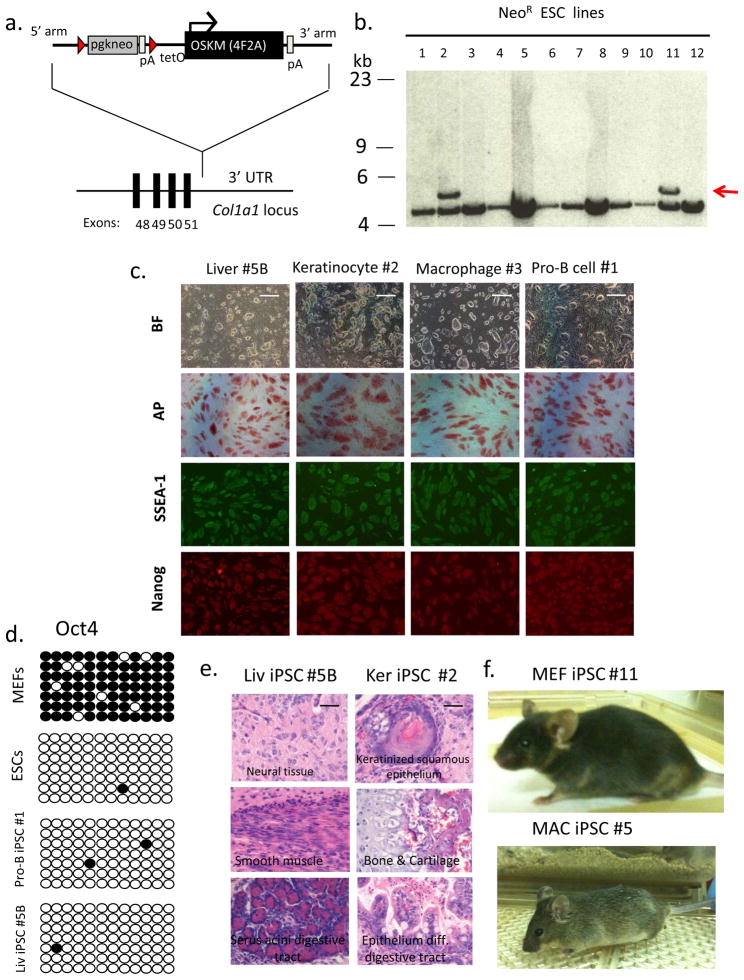
(a) A targeting vector to the 3’ UTR was utilized to deliver four murine reprogramming factors (Oct4, Sox2, Klf4, and c-Myc) as a single dox-inducible polycistronic transgene ,. Red triangles indicate loxP sites. pA indicates poly-adenylation sequence. TetO is tetracycline operator minimal promoter. (b) Southern analysis of DNA obtained from Neo-resistant V19 ES cell colonies was digested with XbaI and probed for correct targeting of the Col1a1 3’ UTR using a 5’ external Col1a1 probe to the genomic sequence outside the targeting vector homology arms . Lines #2 and #11 were correctly targeted as indicated by hybridization to 5.3 kb band (untargeted allele ~ 4.7 kb). (c) Col1a1 4F2A iPS cells express pluripotency markers. Bright field (BF) images and immunostaining for pluripotency markers alkaline phosphatase (AP), SSEA1, and Nanog. Scale bar, 1mm. (d) Oct4 promoter using bisulfite sequencing. Open circles indicate unmethylated and closed circles indicate methylated CpG dinucleotides. Shown are representative sequence analyses from Col1a1 4F2A transgenic MEFs, embryonic stem cells (V6.5), and two Col1a1 4F2A iPSC lines: pro-B-derived #1 and liver-derived line #5B. (e) Hematoxylin and eosin staining of teratomas induced after subcutaneous injection of Col1a1 4F2A Liv iPSC #5B and Ker iPSC #2 into SCID mice indicates Col1a1 4F2A iPSCs contribute to all three germ layers. Scale bar, 100μm (f) Postnatal chimeric mice detected by agouti coat color from Col1a14F2A MEF iPSC #11 and MAC iPSC #5.
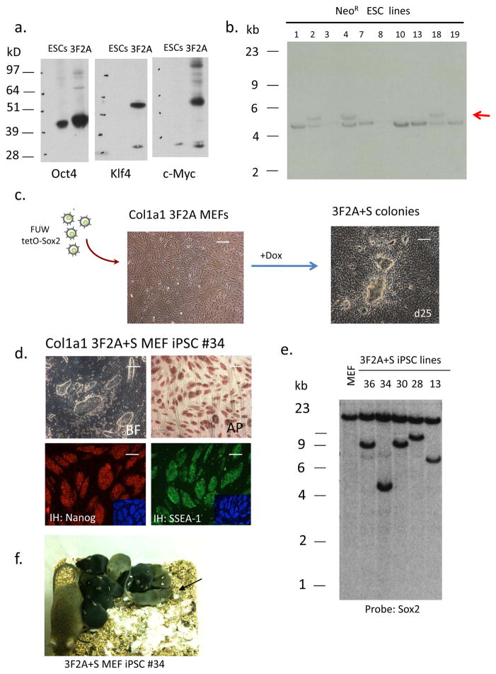
(a) Western blot analysis of 293 cells transiently transfected with pFUW-Ubi 3F2A lentivirus. (b) Southern analysis of DNA obtained from Neo-resistant V19 ES cell colonies. DNA was digested with XbaI and probed for correct targeting using a 5’ Col1a1 probe. Three lines (# 2, 4, 18) were correctly targeted as indicated by hybridization to the 5.3kb band (untargeted allele ~ 4.7 kb). (c) 3F2A MEFs were infected with a Dox-inducible lentivirus carrying a Sox2 cDNA (pFUW-tetO-Sox2) and transferred to ES cell medium containing Dox after 48 hours. Typical iPSC colonies appeared around day 12 and were mechanically passaged at day 25. Scale bar, 1mm. (d) Immunostaining for AP, SSEA1 and Nanog of iPSC line #34. DAPI staining is shown in the inset. Scale bar, 1mm. (e) Southern blot analysis of 3F2A+S iPS cell clones. DNA was isolated from each iPSC line and digested with XbaI. The presence of a single Sox2 proviral copy was detected with a Sox2 cDNA probe for hybridization. A similar sized band was detected in lines #36 and 30 suggesting these cells are sibling subclones from the same infected cell. (f) 3F2A+S MEF derived iPS cells give rise to post-natal chimeras. High contribution chimera (as detected by agouti coat color) obtained after injection of 3F2A+S iPSC line #34 into blastocysts. This chimera contained iPSCs that contributed to the germline as shown by agouti pups present after mating to BDF1 females.
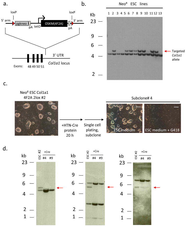
(a) Targeting vector containing loxP sites flanking the 4F2A transgene. A similar targeting scheme as described in Figure 1a was used. (b) Targeting of 3F2A to the Col1a1 locus in R26 M2rtTA transgenic ES cells. Southern analysis of DNA obtained from Neo-resistant ES cell colonies digested with XbaI and probed for correct targeting using a 5’ external Col1a1 probe (compare Fig. 1a). Multiple clones were correctly targeted as indicated by hybridization to the 5.3kb band (untargeted allele ~ 4.7kb). Line #2 was chosen for Cre-induced excision. (c) Recombinant cell-permeable HTN-Cre transduced into Col1a1 2lox 4F2AES cells. Cells were grown in ES cell medium containing HTN-Cre protein for 20 hours. 24 hours later the cells were trypsinized, plated at high dilution (1:10,000) and colonies were picked and expanded before being split into ES medium or ES medium + G418. Bright field images depict a subclone that is G418-sensitive. Scale bars, 1mm. (d) Southern blot analysis of HTN-Cre-induced excision of Col1a1 2lox 4F2A transgene in ESCs. DNA obtained from sister clones of Neo-sensitive ES cell colonies was digested with XbaI and probed using a 5’ external Col1a1 probe (compare Fig. 1a). Two subclones (#4 and 9) do not show hybridization to the ~5.3kb band corresponding to the targeted Col1a1 4F2A after exposure to HTN-Cre protein (left). Oct4 and c-Myc cDNA probes do not hybridize to the expected 5.8kb band (also XbaI digested DNA) corresponding to the Col1a1 4F2A Tg (center and right, respectively).
Comment in
-
Reduce, reuse, reprogram.Nat Methods. 2010 Jan;7(1):39-40. doi: 10.1038/nmeth0110-39. Nat Methods. 2010. PMID: 20038954 No abstract available.
References
-
- Takahashi K, Yamanaka S. Induction of pluripotent stem cells from mouse embryonic and adult fibroblast cultures by defined factors. Cell. 2006;126:663–676. - PubMed
-
- Okita K, Ichisaka T, Yamanaka S. Generation of germline-competent induced pluripotent stem cells. Nature. 2007;448:313–317. - PubMed
-
- Wernig M, et al. In vitro reprogramming of fibroblasts into a pluripotent ES-cell-like state. Nature. 2007;448:318–324. - PubMed
-
- Maherali N, et al. Directly reprogrammed fibroblasts show global epigenetic remodeling and widespread tissue contribution. Cell Stem Cell. 2007;1:55–70. - PubMed
Publication types
MeSH terms
Substances
Grants and funding
LinkOut - more resources
Full Text Sources
Other Literature Sources
Molecular Biology Databases
Research Materials
