

ALDH1A1 is a marker for malignant prostate stem cells and predictor of prostate cancer patients' outcome
- PMID: 20010854
- PMCID: PMC3552330
- DOI: 10.1038/labinvest.2009.127
ALDH1A1 is a marker for malignant prostate stem cells and predictor of prostate cancer patients' outcome
Abstract
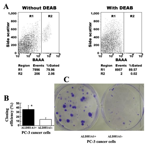
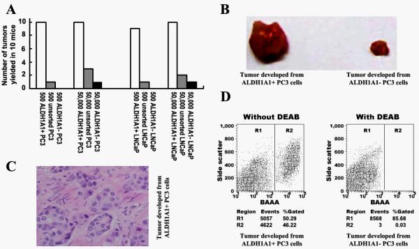
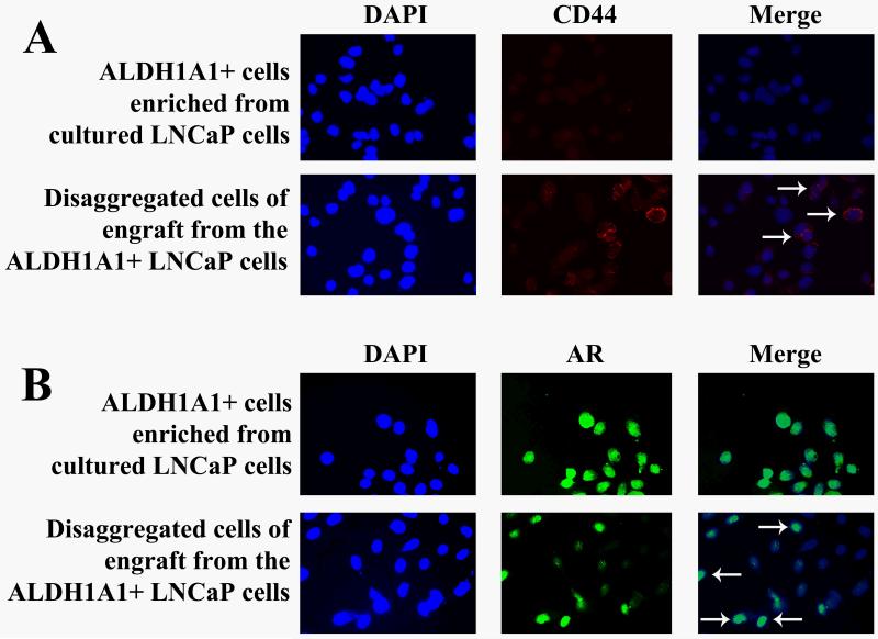
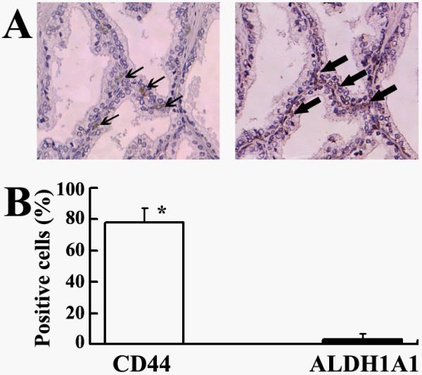
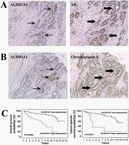
Prostate cancer (PCa) contains a small population of cancer stem cells (CSCs) that contribute to its initiation and progression. The development of specific markers for identification of the CSCs may lead to new diagnostic strategies of PCa. Increased aldehyde dehydrogenase 1A1 (ALDH1A1) activity has been found in the stem cell populations of leukemia and some solid tumors. The aim of the study was to investigate the stem-cell-related function and clinical significance of the ALDH1A1 in human PCa. ALDEFLUOR assay was used to isolate ALDH1A1(+) cells from PCa cell lines. Stem cell characteristics of the ALDH1A1(+) cells were then investigated by in vitro and in vivo approaches. The ALDH1A1 expression was also analyzed by immunohistochemistry in 18 normal prostate and 163 PCa tissues. The ALDH1A1(+) PCa cells showed high clonogenic and tumorigenic capacities, and serially reinitiated transplantable tumors that resembled histopathologic characteristics and heterogeneity of the parental PCa cells in mice. Immunohistochemical analysis of human prostate tissues showed that ALDH1A1(+) cells were sparse and limited to the basal component in normal prostates. However, in tumor specimens, increased ALDH1A1 immunopositivity was found not only in secretory type cancer epithelial cells but also in neuroendocrine tumor populations. Furthermore, the high ALDH1A1 expression in PCa was positively correlated with Gleason score (P=0.01) and pathologic stage (P=0.01), and inversely associated with overall survival and cancer-specific survival of the patients (P=0.00093 and 0.00017, respectively). ALDH1A1 could be a prostate CSC-related marker. Measuring its expression might provide a potential approach to study tumorigenesis of PCa and predict outcome of the disease.
Figures
Similar articles
-
Aldehyde dehydrogenase 1 A1-positive cell population is enriched in tumor-initiating cells and associated with progression of bladder cancer.Cancer Epidemiol Biomarkers Prev. 2010 Feb;19(2):327-37. doi: 10.1158/1055-9965.EPI-09-0865. Cancer Epidemiol Biomarkers Prev. 2010. PMID: 20142235 Free PMC article.
-
ALDH1A1 drives prostate cancer metastases and radioresistance by interplay with AR- and RAR-dependent transcription.Theranostics. 2024 Jan 1;14(2):714-737. doi: 10.7150/thno.88057. eCollection 2024. Theranostics. 2024. PMID: 38169509 Free PMC article.
-
Increased Expression of ALDH1A1 in Prostate Cancer is Correlated With Tumor Aggressiveness: A Tissue Microarray Study of Iranian Patients.Appl Immunohistochem Mol Morphol. 2017 Sep;25(8):592-598. doi: 10.1097/PAI.0000000000000343. Appl Immunohistochem Mol Morphol. 2017. PMID: 26894647
-
Aldehyde dehydrogenase 1A1 in stem cells and cancer.Oncotarget. 2016 Mar 8;7(10):11018-32. doi: 10.18632/oncotarget.6920. Oncotarget. 2016. PMID: 26783961 Free PMC article. Review.
-
Lower RNA expression of ALDH1A1 distinguishes the favorable risk group in acute myeloid leukemia.Mol Biol Rep. 2022 Apr;49(4):3321-3331. doi: 10.1007/s11033-021-07073-7. Epub 2022 Jan 14. Mol Biol Rep. 2022. PMID: 35028852 Review.
Cited by
-
Pathobiological implications of the expression of EGFR, pAkt, NF-κB and MIC-1 in prostate cancer stem cells and their progenies.PLoS One. 2012;7(2):e31919. doi: 10.1371/journal.pone.0031919. Epub 2012 Feb 23. PLoS One. 2012. PMID: 22384099 Free PMC article.
-
ASPM promotes prostate cancer stemness and progression by augmenting Wnt-Dvl-3-β-catenin signaling.Oncogene. 2019 Feb;38(8):1340-1353. doi: 10.1038/s41388-018-0497-4. Epub 2018 Sep 28. Oncogene. 2019. PMID: 30266990
-
Role of prostate cancer stem-like cells in the development of antiandrogen resistance.Cancer Drug Resist. 2022 Jun 1;5(2):459-471. doi: 10.20517/cdr.2022.07. eCollection 2022. Cancer Drug Resist. 2022. PMID: 35800367 Free PMC article. Review.
-
Aldehyde Dehydrogenase 1B1 as a Modulator of Pancreatic Adenocarcinoma.Pancreas. 2016 Jan;45(1):117-22. doi: 10.1097/MPA.0000000000000542. Pancreas. 2016. PMID: 26566217 Free PMC article.
-
Cancer stem cells and chemoresistance: The smartest survives the raid.Pharmacol Ther. 2016 Apr;160:145-58. doi: 10.1016/j.pharmthera.2016.02.008. Epub 2016 Feb 17. Pharmacol Ther. 2016. PMID: 26899500 Free PMC article. Review.
References
-
- Cookson MS, Aus G, Burnett AL, et al. Variation in the definition of biochemical recurrence in patients treated for localized prostate cancer: the American Urological Association Prostate Guidelines for Localized Prostate Cancer Update Panel report and recommendations for a standard in the reporting of surgical outcomes. J Urol. 2007;177:540–5. - PubMed
-
- Reya T, Morrison SJ, Clarke MF, et al. Stem cells, cancer, and cancer stem cells. Nature. 2001;414:105–11. - PubMed
-
- Jordan CT, Guzman ML, Noble M. Cancer stem cells. N Engl J Med. 2006;355:1253–61. - PubMed
Publication types
MeSH terms
Substances
Grants and funding
LinkOut - more resources
Full Text Sources
Medical
Miscellaneous
