

The promyelocytic leukemia zinc-finger gene, PLZF, is frequently downregulated in malignant mesothelioma cells and contributes to cell survival
- PMID: 20010871
- PMCID: PMC2842080
- DOI: 10.1038/onc.2009.455
The promyelocytic leukemia zinc-finger gene, PLZF, is frequently downregulated in malignant mesothelioma cells and contributes to cell survival
Abstract
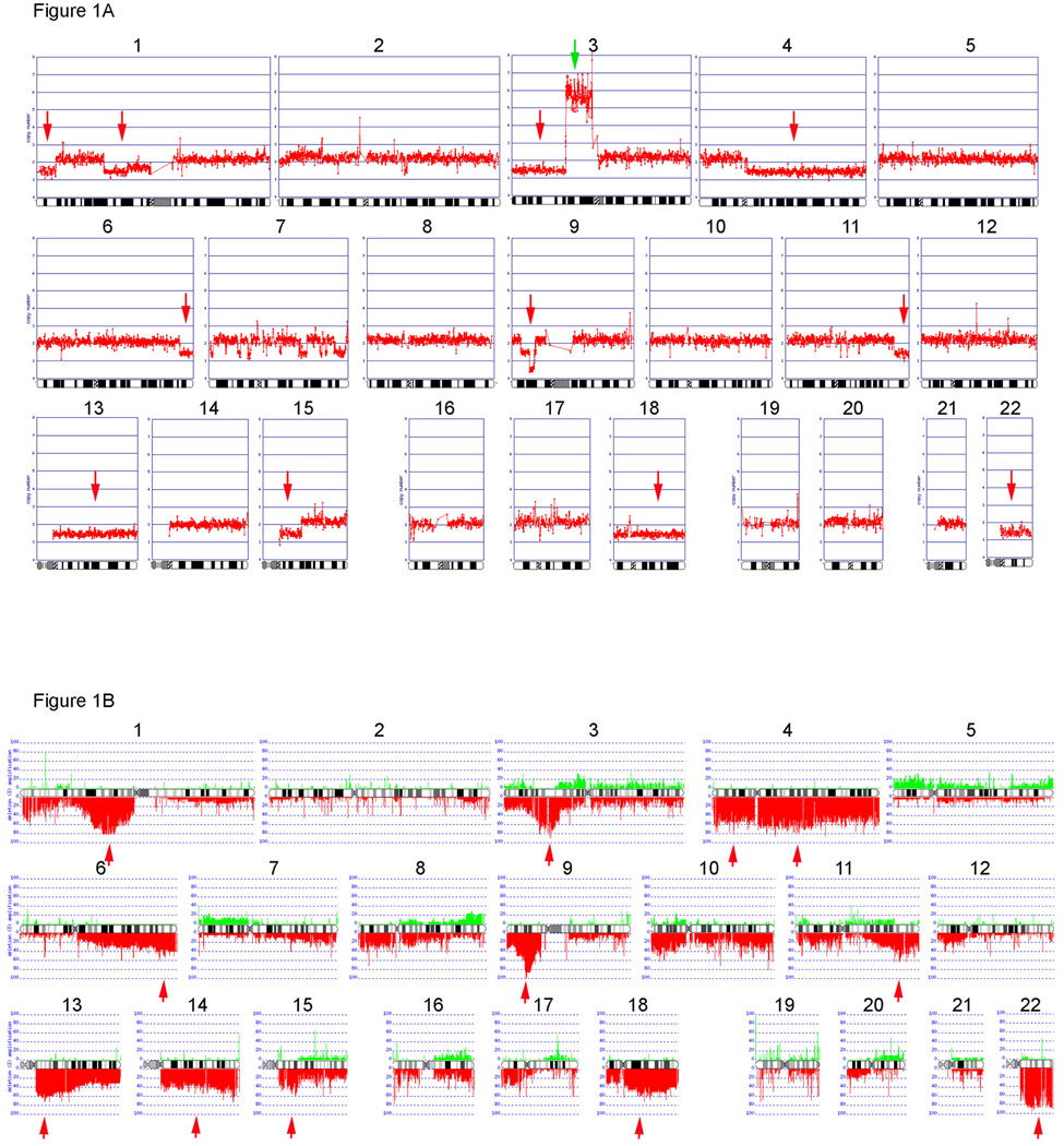
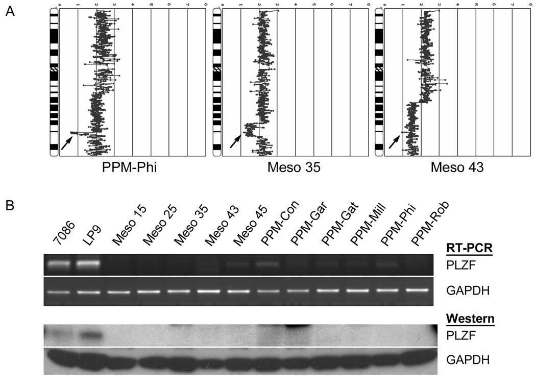
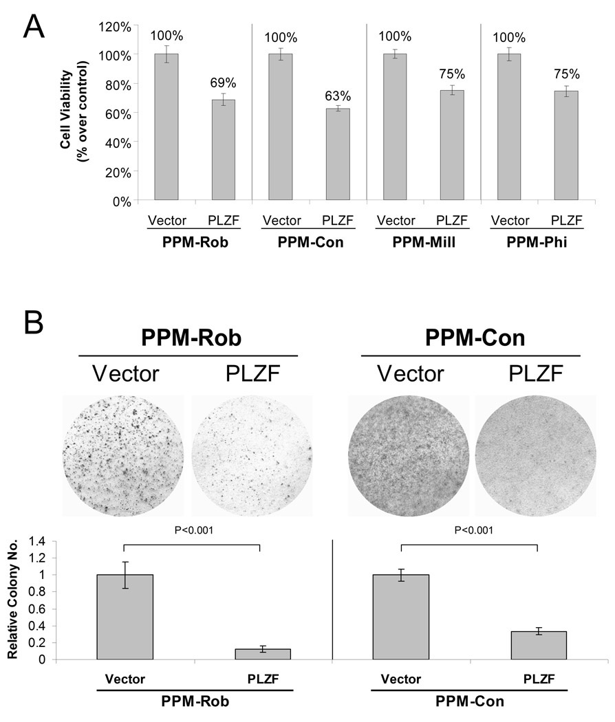
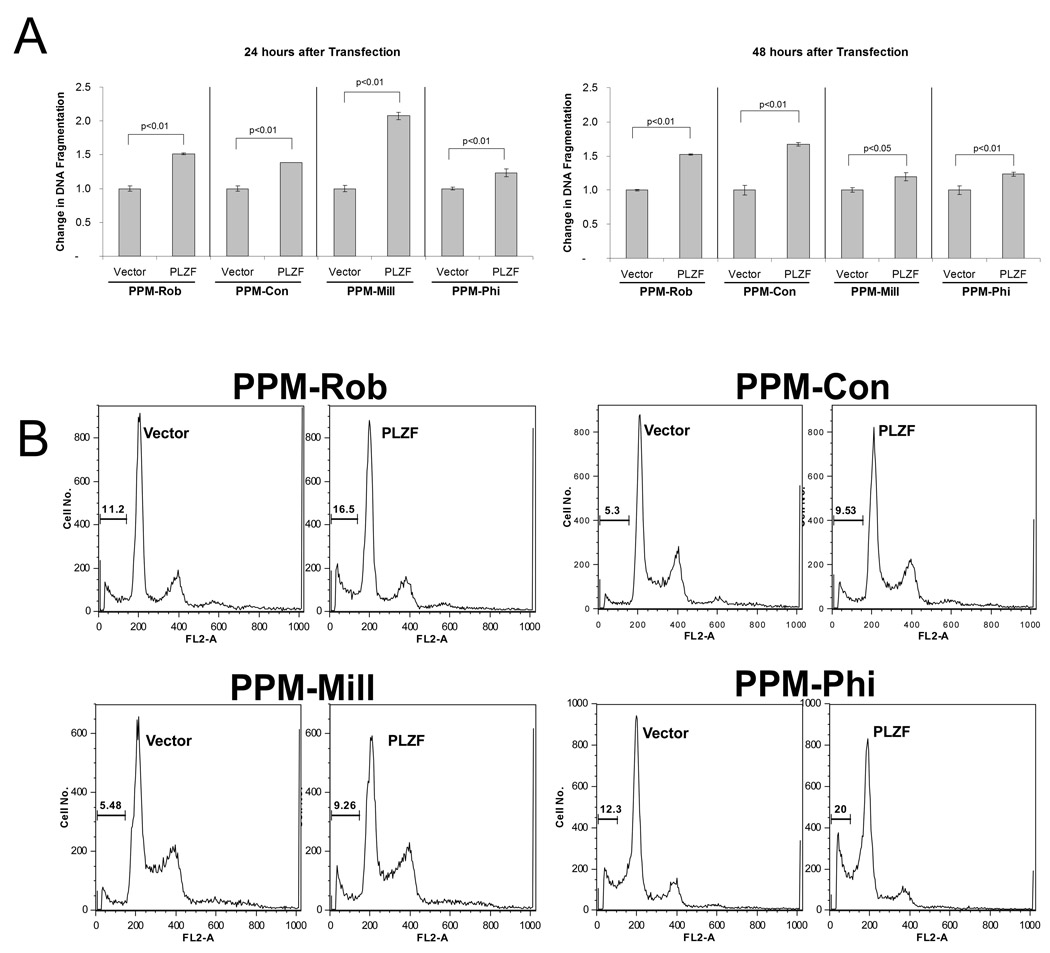
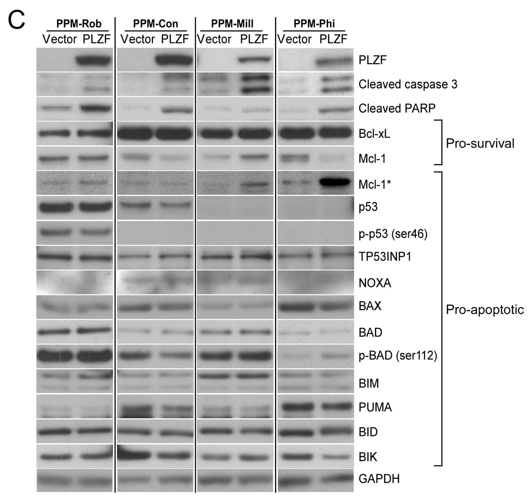
DNA copy number analysis was performed, using single-nucleotide polymorphism mapping arrays, to fine map genomic imbalances in human malignant mesothelioma (MM) cell lines derived from primary tumors. Chromosomal losses accounted for the majority of genomic imbalances. All 22 cell lines examined showed homozygous deletions of 9p21.3, centering at the CDKN2A/ARF and CDKN2B loci. Other commonly underrepresented segments included 1p36, 1p22, 3p21-22, 4q13, 4q34, 11q23, 13q12-13, 14q32, 15q15, 18q12, and 22q12, each observed in 55-90% of cell lines. Focal deletions of 11q23 encompassed the transcriptional repressor gene promyelocytic leukemia zinc finger (PLZF), which was validated by analysis of genomic DNA using real-time polymerase chain reaction (PCR). Semi-quantitative RT-PCR and immunoblot analysis revealed that PLZF is greatly downregulated in MM cell lines compared with non-malignant mesothelial cells. Ectopic expression of PLZF in PLZF-deficient MM cells resulted in decreased cell viability, reduced colony formation, as well as increased apoptosis, the latter based on results of various cell death assays and the observation of increased cleavage of caspase 3, PARP, and Mcl-1. These data indicate that deletions of PLZF are a common occurrence in MM and that downregulation of PLZF may contribute to MM pathogenesis by promoting cell survival.
Conflict of interest statement
The authors declare no conflict of interest.
Figures
Similar articles
-
Fine-mapping loss of gene architecture at the CDKN2B (p15INK4b), CDKN2A (p14ARF, p16INK4a), and MTAP genes in head and neck squamous cell carcinoma.Arch Otolaryngol Head Neck Surg. 2006 Apr;132(4):409-15. doi: 10.1001/archotol.132.4.409. Arch Otolaryngol Head Neck Surg. 2006. PMID: 16618910
-
Deregulated expression of promyelocytic leukemia zinc finger protein in B-cell chronic lymphocytic leukemias does not affect cyclin A expression.Hematol J. 2000;1(1):15-27. doi: 10.1038/sj.thj.6200012. Hematol J. 2000. PMID: 11920165
-
CDKN2A, CDKN2B, and MTAP gene dosage permits precise characterization of mono- and bi-allelic 9p21 deletions in childhood acute lymphoblastic leukemia.Genes Chromosomes Cancer. 2003 May;37(1):44-57. doi: 10.1002/gcc.10188. Genes Chromosomes Cancer. 2003. PMID: 12661005
-
The Yin and Yang-Like Clinical Implications of the CDKN2A/ARF/CDKN2B Gene Cluster in Acute Lymphoblastic Leukemia.Genes (Basel). 2021 Jan 9;12(1):79. doi: 10.3390/genes12010079. Genes (Basel). 2021. PMID: 33435487 Free PMC article. Review.
-
Role of promyelocytic leukemia zinc finger protein in proliferation of cultured human corneal endothelial cells.Cornea. 2007 Oct;26(9 Suppl 1):S55-8. doi: 10.1097/ICO.0b013e31812f6b67. Cornea. 2007. PMID: 17881917 Review.
Cited by
-
PLZF mediates the PTEN/AKT/FOXO3a signaling in suppression of prostate tumorigenesis.PLoS One. 2013 Dec 10;8(12):e77922. doi: 10.1371/journal.pone.0077922. eCollection 2013. PLoS One. 2013. PMID: 24339862 Free PMC article.
-
Targeting LRRC15 Inhibits Metastatic Dissemination of Ovarian Cancer.Cancer Res. 2022 Mar 15;82(6):1038-1054. doi: 10.1158/0008-5472.CAN-21-0622. Cancer Res. 2022. PMID: 34654724 Free PMC article.
-
Malignant pleural mesothelioma: an update.J Bras Pneumol. 2021 Dec 13;47(6):e20210129. doi: 10.36416/1806-3756/e20210129. eCollection 2021. J Bras Pneumol. 2021. PMID: 34909922 Free PMC article. Review.
-
Understanding PLZF: two transcriptional targets, REDD1 and smooth muscle α-actin, define new questions in growth control, senescence, self-renewal and tumor suppression.Cell Cycle. 2011 Mar 1;10(5):771-5. doi: 10.4161/cc.10.5.14829. Epub 2011 Mar 1. Cell Cycle. 2011. PMID: 21311223 Free PMC article.
-
The Promyelocytic Leukemia Zinc Finger Transcription Factor Is Critical for Human Endometrial Stromal Cell Decidualization.PLoS Genet. 2016 Apr 1;12(4):e1005937. doi: 10.1371/journal.pgen.1005937. eCollection 2016 Apr. PLoS Genet. 2016. PMID: 27035670 Free PMC article.
References
-
- Altomare DA, Vaslet CA, Skele KL, De Rienzo A, Devarajan K, Jhanwar SC, et al. A mouse model recapitulating molecular features of human mesothelioma. Cancer Res. 2005;65:8090–8095. - PubMed
-
- Balsara BR, Bell DW, Sonoda G, De Rienzo A, du Manoir S, Jhanwar S, et al. Comparative genomic hybridization and loss of heterozygosity analyses identify a common region of deletion at 15q11.1-15 in human malignant mesothelioma. Cancer Res. 1999;59:450–454. - PubMed
-
- Barna M, Merghoub T, Costoya JA, Ruggero D, Branford M, Bergia A, et al. Plzf mediates transcriptional repression of HoxD gene expression through chromatin remodeling. Dev Cell. 2002;3:499–510. - PubMed
-
- Bernardo MV, Yelo E, Gimeno L, Campillo JA, Parrado A. Identification of apoptosis-related PLZF target genes. Biochem Biophys Res Commun. 2007;359:317–322. - PubMed
Publication types
MeSH terms
Substances
Grants and funding
LinkOut - more resources
Full Text Sources
Research Materials
Miscellaneous
