. 2010 Jan 19;102(2):351-60.
doi: 10.1038/sj.bjc.6605486. Epub 2009 Dec 15.

Hypoxia potentiates Notch signaling in breast cancer leading to decreased E-cadherin expression and increased cell migration and invasion

Affiliations

Hypoxia potentiates Notch signaling in breast cancer leading to decreased E-cadherin expression and increased cell migration and invasion

J Chen et al. Br J Cancer. .

Abstract

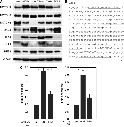
Background: Epithelial-to-mesenchymal transition (EMT) is associated with decreased adhesion and acquisition of metastatic potential of breast cancer cells. Epithelial-to-mesenchymal transition is mediated, in part, by two transcription repressors, Snail and Slug, that are known to be targets of the Notch signaling pathway, and JAGGED1-induced Notch activation increases EMT. However, the events that lead to increased Notch activity during EMT of breast cancer cells are unknown.

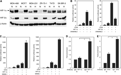
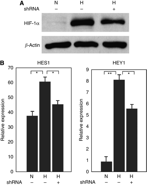
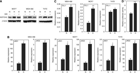
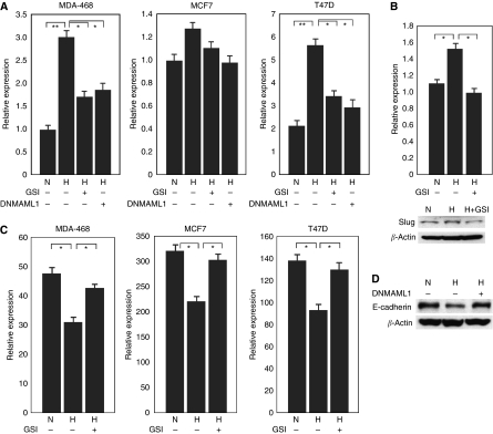
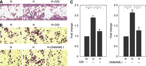
Methods: The accumulation of hypoxia inducible factors (HIFs) under hypoxia was detected by western blot analysis, and their effects on Notch signaling were measured by an in vitro Notch reporter assay. The expression of Notch target genes under hypoxia was tested by real-time PCR. The knockdown of HIF-1alpha was mediated by retroviral delivery of shRNA. The expression of Slug and Snail under hypoxia was measured by real-time PCR. Breast cancer cell migration and invasion under hypoxia were tested with cell migration and invasion kits.

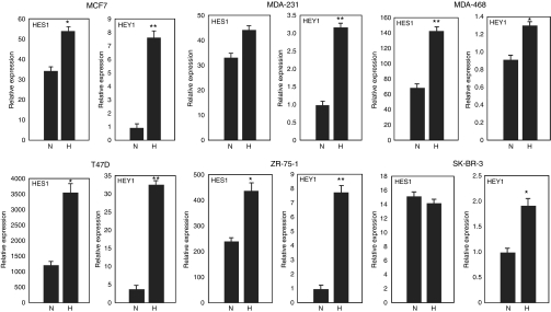
Results: Hypoxia increased the expression of Notch target genes such as HES1 and HEY1 in breast cancer cells, as was expression of Notch receptors and ligands. The mechanism is likely to involve the accumulation of HIF-1alpha and HIF-2alpha in these cells by hypoxia, which synergised with the Notch co-activator MAML1 in potentiating Notch activity. Hypoxia inducible factor-1alpha was found to bind to HES1 promoter under hypoxia. Knockdown of HIF-1alpha with shRNA inhibited both HES1 and HEY1 expression under hypoxia. Hypoxia increased the expression of Slug and Snail, and decreased the expression of E-cadherin, hallmarks of EMT. Notch pathway inhibition abrogated the hypoxia-mediated increase in Slug and Snail expression, as well as decreased breast cancer cell migration and invasion.

Conclusion: Hypoxia-mediated Notch signaling may have an important role in the initiation of EMT and subsequent potential for breast cancer metastasis.


Figures

Figure 1
Figure 1
Notch signaling is active in human breast cancer cells. (A) Expression of Notch receptors, ligands and Notch target gene HES1 in breast cancer cells as detected by western blot analysis with specific antibodies. 468: MDA-468 cells; 231: MDA-231 cells. (B) Part of human HES1 5’ upstream sequence. The RBP-Jκ binding sites are capitalised. The primer sequences flanking the RBP-Jκ binding sites are underlined. The control primer sequences at approximately 3.7-kb upstream are also underlined. (C) Fold enrichment of the binding of NOTCH3 intracellular domain or MAML1 to the RBP-Jκ binding sites of human HES1 promoter in MCF7 cells with or without GSI treatment. Chromatin immunoprecipitation (CHIP) was performed with either normal IgG, ICN3-specific antibody or MAML1 antibody followed by real-time PCR with primers flanking the RBP-Jκ binding sites of human HES1 promoter. *P<0.01; **P<0.05
Figure 2
Figure 2
Hypoxia inducible factor (HIF)-1α and HIF-2α accumulated in breast cancer cells with hypoxia and they potentiated Notch signaling. (A) Western blot analysis of HIF-1α (the upper band marked by the arrow) and HIF-2α expression in breast cancer cells under 21% O2 or 1% O2 culture conditions for 24 h. H, hypoxia; N, normoxia. (B) Enhanced ICN3 signaling by HIF-1α or HIF-2α as determined by an in vitro Notch reporter assay, which was inhibited by dominant negative MAML1. (C) Hypoxia inducible factor (HIF)-1α and HIF-2α synergise with MAML1 in potentiating ICN3-mediated Notch signaling. (D) Enrichment of HIF-1α at HES1 promoter by hypoxia in MCF7 cells. Fold enrichment of the binding of HIF-1α to the RBP-Jκ binding sites of human HES1 promoter in MCF7 cells under 1% O2 culture condition. Normal IgG or HIF-1α-specific antibodies were used for CHIP experiments followed by real-time PCR with primers flanking the RBP-Jκ binding sites of HES1 promoter. H, hypoxia; N, normoxia; *P<0.01. (E) Increased binding of NOTCH3 intracellular domain to the RBP-Jκ binding sites of HES1 promoter in MCF7 cells under hypoxia. H: hypoxia; N, normoxia; *P<0.05.
Figure 3
Figure 3
Increased Notch target gene expression in breast cancer cells by hypoxia. Real-time PCR analysis of the expression of Notch target genes HES1 and HEY1 in breast cancer cells under 1% O2 culture condition for 24 h. For MCF7 cells: *P<0.05, **P<0.001. For MDA-231 cells: **P<0.01. For MDA-468 cells: *P<0.05, **P<0.01. For T47D cells: *P<0.01, **P<0.001. For ZR-75-1 cells: *P<0.05, **P<0.001. For SK-BR-3 cells: *P<0.05. H, hypoxia; N, normoxia.
Figure 4
Figure 4
The increased expression of HES1 and HEY1 in MCF7 cells under hypoxia is HIF-1α dependant. (A) Western blot analysis of HIF-1α expression in MCF7 cells under hypoxia with the knockdown of HIF-1α by shRNA. (B) Real-time PCR analysis of HES1 and HEY1 expression in MCF7 cells under hypoxia with the knockdown of HIF-1α by shRNA. *P<0.05, **P<0.001. H, hypoxia; N, normoxia.
Figure 5
Figure 5
Increased Notch receptor and Notch ligand expression in breast cancer cells by hypoxia. (A) Western blot analysis of the expression of NOTCH3 in breast cancer cells under 21% O2 or 1% O2 culture conditions with or without GSI treatment. H, hypoxia; N, normoxia. (B) Expression of Notch ligands JAGGED1 and JAGGED2 in breast cancer cells under hypoxia. *P<0.01. (C) Expression of Notch ligand DELTA1 in breast cancer cells under hypoxia. *P<0.05; **P<0.01. (D) MCF7 cells were transfected with HES1-luc and TK-Renilla luciferase reporter genes and were cultured at 21% or 1% O2 conditions. Dual-luciferase assays were performed 48 h after transfection. *P<0.05; H, hypoxia; N, normoxia.
Figure 6
Figure 6
Altered expression of Slug, Snail and E-cadherin in breast cancer cells by hypoxia. (A) Real-time PCR analysis of the expression of Snail in breast cancer cells with hypoxia. The increased expression of Snail with hypoxia was abrogated by either GSI treatment or DNMAML1. *P<0.05; **P<0.01. (B) Slug expression (both mRNA level and protein level) was increased in MDA-231 cells with hypoxia, which was abrogated by GSI treatment. *P<0.05. (C) Real-time PCR analysis of the expression of E-cadherin in breast cancer cells under 21% O2 or 1% O2 culture conditions with or without GSI treatment. *P<0.05. (D) Western blot analysis of E-cadherin expression in MCF7 cells with hypoxia. Dominant negative MAML1 abrogated the downregulation of E-cadherin during hypoxia. H, hypoxia; N, normoxia.
Figure 7
Figure 7
Notch signaling is required for hypoxia-mediated MDA-468 cell migration and invasion. (A) The closure of scratch wound of MDA-468 cells was facilitated by hypoxia and was inhibited by GSI treatment. (B and C) The migration of MDA-468 breast cancer cells across a basement membrane toward the serum attraction was elevated by hypoxia and was abrogated by either GSI treatment or DNMAML1. *P<0.05; H, hypoxia; N, normoxia.

References

    1. Ayyanan A, Civenni G, Ciarloni L, Morel C, Mueller N, Lefort K, Mandinova A, Raffoul W, Fiche M, Dotto GP, Brisken C (2006) Increased Wnt signaling triggers oncogenic conversion of human breast epithelial cells by a Notch-dependent mechanism. Proc Natl Acad Sci USA 103: 3799–3804 - PMC - PubMed
    1. Bedogni B, Warneke JA, Nickoloff BJ, Giaccia AJ, Powell MB (2008) Notch1 is an effector of Akt and hypoxia in melanoma development. J Clin Invest 118: 3660–3670 - PMC - PubMed
    1. Callahan R, Egan SE (2004) Notch signaling in mammary development and oncogenesis. J Mammary Gland Biol Neoplasia 9: 145–163 - PubMed
    1. Chen Y, De Marco MA, Graziani I, Gazdar AF, Strack PR, Miele L, Bocchetta M (2007) Oxygen concentration determines the biological effects of NOTCH-1 signaling in adenocarcinoma of the lung. Cancer Res 67: 7954–7959 - PubMed
    1. Dievart A, Beaulieu N, Jolicoeur P (1999) Involvement of Notch1 in the development of mouse mammary tumors. Oncogene 18: 5973–5981 - PubMed

Publication types

MeSH terms