. 2009 Dec 9;4(12):e8234.
doi: 10.1371/journal.pone.0008234.

Lymphatic vascularisation and involvement of Lyve-1+ macrophages in the human onchocerca nodule

Affiliations

Lymphatic vascularisation and involvement of Lyve-1+ macrophages in the human onchocerca nodule

Tarik Attout et al. PLoS One. .

Abstract

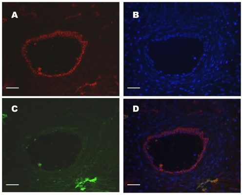
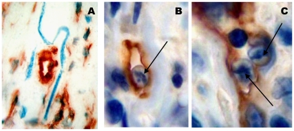
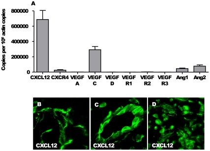
Onchocerciasis, caused by the filarial nematode Onchocerca volvulus, is a parasitic disease leading to debilitating skin disease and blindness, with major economic and social consequences. The pathology of onchocerciasis is principally considered to be a consequence of long-standing host inflammatory responses. In onchocerciasis a subcutaneous nodule is formed around the female worms, the core of which is a dense infiltrate of inflammatory cells in which microfilariae are released. It has been established that the formation of nodules is associated with angiogenesis. In this study, we show using specific markers of endothelium (CD31) and lymphatic endothelial cells (Lyve-1, Podoplanin) that not only angiogenesis but also lymphangiogenesis occurs within the nodule. 7% of the microfilariae could be found within the lymphatics, but none within blood vessels in these nodules, suggesting a possible route of migration for the larvae. The neovascularisation was associated with a particular pattern of angio/lymphangiogenic factors in nodules of onchocerciasis patients, characterized by the expression of CXCL12, CXCR4, VEGF-C, Angiopoietin-1 and Angiopoietin-2. Interestingly, a proportion of macrophages were found to be positive for Lyve-1 and some were integrated into the endothelium of the lymphatic vessels, revealing their plasticity in the nodular micro-environment. These results indicate that lymphatic as well as blood vascularization is induced around O. volvulus worms, either by the parasite itself, e.g. by the release of angiogenic and lymphangiogenic factors, or by consecutive host immune responses.


Conflict of interest statement

Competing Interests: The authors have declared that no competing interests exist.

Figures

Figure 1
Figure 1. Blood and lymphatic identification in onchocercoma.
Five-micrometer serial sections were stained with (A) CD31 or (C) Lyve-1; (B) DAPI nuclear counterstaining. (D) Merging of A, B and C. Number of analysed nodules = 5. Scale: A, B, C, D: bar = 25 µm.
Figure 2
Figure 2. Vascular organisation of onchocercoma.
(A) The different zones in nodules: zone A = the outer fibrovascular capsule (black stars), zone B = the inner adult worm bundle with surrounding extracellular matrix interspersed with solitary cells (white stars), zone C = a dense cellular infiltrate surrounding and in contact with the worm (blue stars). (B–M) The blood and lymphatic system in the nodules: sections of onchocerca nodules were stained for CD31 (B, E, H, K) to detect blood and lymphatic vessels, in the zone A (B), in the zone B (E), in the zone C (H), close to a worm section (K) and for Podoplanin (C, F, I, L) in the zone A (C), in the zone B (F), in the zone C (I), close to a worm section (L) or for Lyve-1 (D, G, J, M) in the zone A (D), in the zone B (G), in the zone C (J), close to a worm section (M), to identify lymphatics vessels. Number of analysed nodules = 10. O.v.: O. volvulus section. Scales: A, bar = 300 µm; B,C,D,E,G,J,K,M bar = 25 µm; F,H,I,L, bar = 10 µm.
Figure 3
Figure 3. Presence of microfilariae in lymphatic vessels from the nodule.
(A) Microfilariae (blue turquoise) in nodule close to a lymphatic section. (B & C) Sections of microfilariae (arrows) within lymphatic vessels in the nodules (Lyve-1 staining). Number of analysed nodules = 10. Scales: A, bar = 20 µm; B, C, bar = 10 µm.
Figure 4
Figure 4. Localisation of microfilariae in the dermis of onchocercian patients.
(A) Lyve-1 staining showing two dilated lymphatic vessels (black arrow)on a paraffin section (B) Lyve-1 staining revealing one dilated lymphatic vessel with mononuclear cells on a cryosection (C) Podoplanin staining showing a collapsed lymphatic vessel on a cryosection. (D) Microfilaria (white arrow) section in a perivascular space near to a lymphatic vessel. Number of analysed skin snips = 8. Scales: A,B,C,D, bar = 50 µm.
Figure 5
Figure 5. Angio- and lymphangiogenic markers in nodules.
(A) A particular pattern of angio/lymphangiogenic factors was established by qPCR in nodules of onchocerciasis patients, characterized by the significant expression of CXCL12, CXCR4, VEGF-C, Angiopoietin-1 and Angiopoietin-2. Number of analysed nodules = 33. Data were analyzed using Prism version 4.0 (GraphPad) and presented as mean±SEM. (B-C-D) Expression of CXCL12 in nodule by immunofluorescence. Scales: B, bar = 20 µm.
Figure 6
Figure 6. Lyve-1+ macrophages in nodule.
(A) Individual Lyve-1+ mononuclear cells in the fibrous capsule of nodules. (B) Detail of a Lyve-1+ mononuclear cell phagocytising apoptotic neutrophil. (C–K) Double Immunostaining for Lyve-1/CD68 to establish the macrophage lineage of Lyve-1+ mononuclear cells: (C & F) Lyve-1+ mononuclear cells, (D & G) CD68+ macrophage, (E & H) merging C & D and F & G. (I–K) Incorporation of Lyve-1+ macrophages into lymphatics: (I) Lyve-1+ lymphatic vessel, (J) CD68+ macrophage, (K) merging I & J. Number of analysed nodules = 10. Scales: A,B,F,G,H, bar = 50 µm.

Similar articles

Cited by

References

    1. Beveridge I, Kummerow EL, Wilkinson P. Observations on Onchocerca gibsoni and nodule development in naturally-infected cattle in Australia. Tropenmed Parasitol. 1980;31:75–81. - PubMed
    1. Bain O, Bussieras J, Amegee E. [Further description of two African bovine's Onchocerca (author's transl)]. Ann Parasitol Hum Comp. 1976;51:461–471. - PubMed
    1. Collins RC, Lujan R, Figueroa H, Campbell CC. Early formation of the nodule in Guatemalan onchocerciasis. Am J Trop Med Hyg. 1982;31:267–269. - PubMed
    1. Brattig NW. Pathogenesis and host responses in human onchocerciasis: impact of Onchocerca filariae and Wolbachia endobacteria. Microbes Infect. 2004;6:113–128. - PubMed
    1. Allen JE, Adjei O, Bain O, Hoerauf A, Hoffmann WH, et al. Of mice, cattle, and humans: the immunology and treatment of river blindness. PLoS Negl Trop Dis. 2008;2:e217. - PMC - PubMed

Publication types

MeSH terms

Substances