. 2009 Dec 9;4(12):e8221.
doi: 10.1371/journal.pone.0008221.

Glimepiride reduces the expression of PrPc, prevents PrPSc formation and protects against prion mediated neurotoxicity in cell lines

Affiliations

Glimepiride reduces the expression of PrPc, prevents PrPSc formation and protects against prion mediated neurotoxicity in cell lines

Clive Bate et al. PLoS One. .

Abstract

Background: A hallmark of the prion diseases is the conversion of the host-encoded cellular prion protein (PrP(C)) into a disease related, alternatively folded isoform (PrP(Sc)). The accumulation of PrP(Sc) within the brain is associated with synapse loss and ultimately neuronal death. Novel therapeutics are desperately required to treat neurodegenerative diseases including the prion diseases.

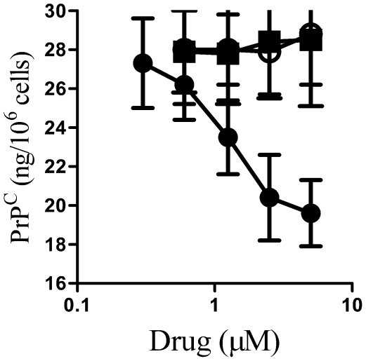
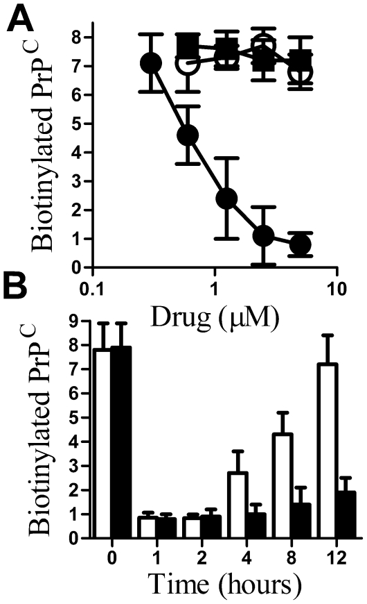
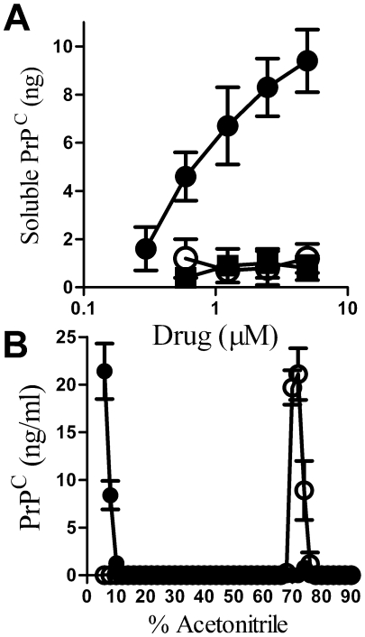
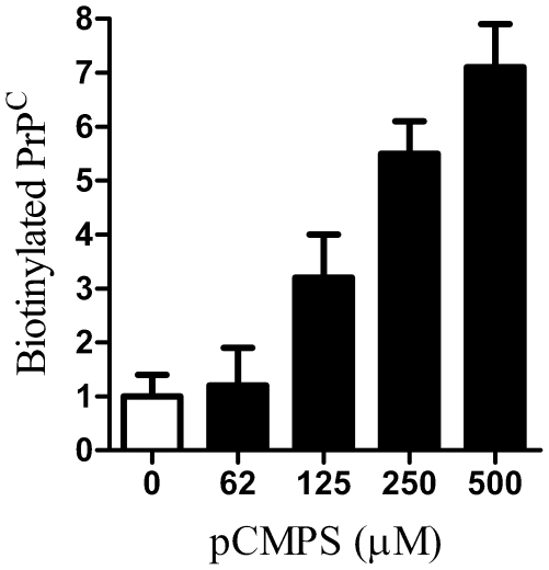
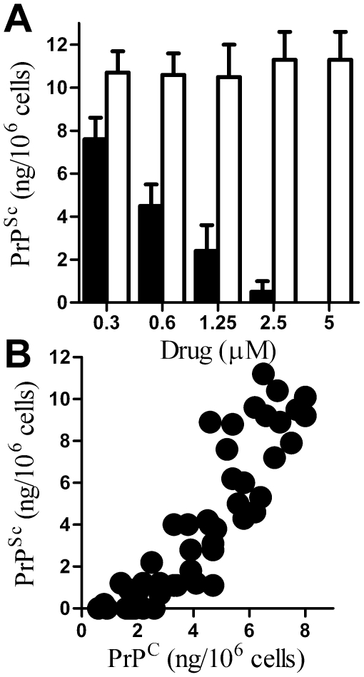
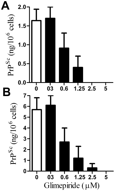
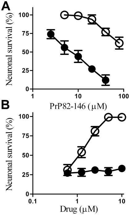
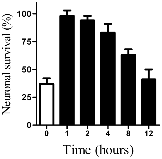
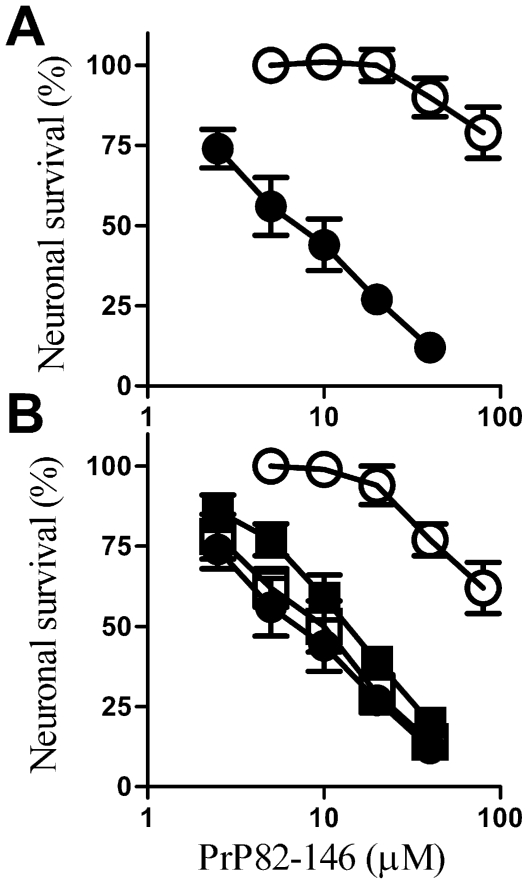
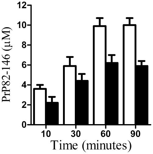
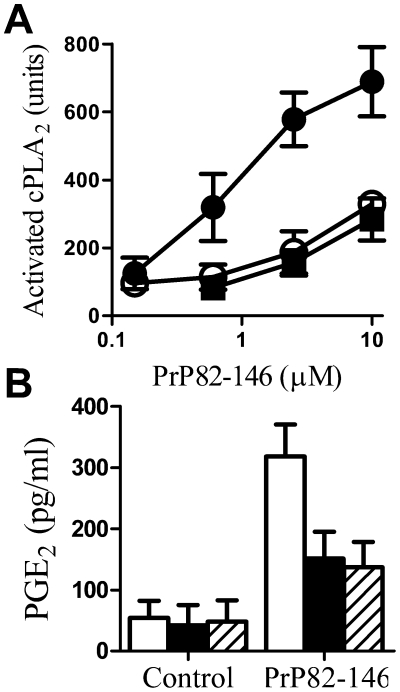
Principal findings: Treatment with glimepiride, a sulphonylurea approved for the treatment of diabetes mellitus, induced the release of PrP(C) from the surface of prion-infected neuronal cells. The cell surface is a site where PrP(C) molecules may be converted to PrP(Sc) and glimepiride treatment reduced PrP(Sc) formation in three prion infected neuronal cell lines (ScN2a, SMB and ScGT1 cells). Glimepiride also protected cortical and hippocampal neurones against the toxic effects of the prion-derived peptide PrP82-146. Glimepiride treatment significantly reduce both the amount of PrP82-146 that bound to neurones and PrP82-146 induced activation of cytoplasmic phospholipase A(2) (cPLA(2)) and the production of prostaglandin E(2) that is associated with neuronal injury in prion diseases. Our results are consistent with reports that glimepiride activates an endogenous glycosylphosphatidylinositol (GPI)-phospholipase C which reduced PrP(C) expression at the surface of neuronal cells. The effects of glimepiride were reproduced by treatment of cells with phosphatidylinositol-phospholipase C (PI-PLC) and were reversed by co-incubation with p-chloromercuriphenylsulphonate, an inhibitor of endogenous GPI-PLC.

Conclusions: Collectively, these results indicate that glimepiride may be a novel treatment to reduce PrP(Sc) formation and neuronal damage in prion diseases.


Conflict of interest statement

Competing Interests: The authors have declared that no competing interests exist.

Figures

Figure 1
Figure 1. Glimepiride reduced neuronal PrPC expression.
The amount of PrPC in whole cell extracts from primary cortical neurones treated for 1 hour with different concentrations of glimepiride (•), glibenclamide (○) or glipizide (▪) as shown. Values shown are the mean average amount of PrPC in neuronal extracts (ng/106 cells) ± SD, n = 12.
Figure 2
Figure 2. Glimepirde reduced the amount of PrPC at the surface of neurones.
(A) The amount of cell surface PrPC on neurones treated for 1 hour with different concentrations of glimepiride (•), glibenclamide (○) or glipizide (▪) as shown. Values shown are the mean average amount of biotinylated PrPC in cell extracts (ng/106 cells) ± SD, n = 9. (B) The amount of cell surface PrPC in cell extracts from cortical neurones taken at different time points after the addition of 5 µM glimepiride alone () or a mixture containing 5 µM glimepiride and 20 µg/ml cycloheximide (▪) as shown. Values shown are the mean average amount of biotinylated PrPC in cell extracts (ng/106 cells) ± SD, n = 9.
Figure 3
Figure 3. Glimepiride released PrPC from cortical neurones.
(A) The amount of PrPC in the supernatant of cortical neurones treated for 1 hour with different concentrations of glimepiride (•), glibenclamide (○) or glipizide (▪). Values shown are the mean average amount of soluble PrPC (ng/106 cells) ± SD, n = 9. (B) The amount of PrPC molecules eluted from C18 columns following elution with a gradient of acetonitrile in water containing 0.1% TFA. Fractions eluted from C18 columns loaded with whole cell extracts (○) or with supernatants from cortical neurones treated with 5 µM glimepiride (•). Values shown are the mean average amount of PrPC (ng/ml) ± SD, n = 6.
Figure 4
Figure 4. Glimepiride induced release of PrPC is reversed by p-CMPS.
The amount of PrPC at the surface of cortical neurones treated for 1 hour with 5 µM glimepiride alone (□) or with combinations containing 5 µM glimepiride and different concentrations of p-CMPS as shown (▪). Values shown are the mean average amount of biotinylated-PrPC (ng/106 cells) ± SD, n = 8.
Figure 5
Figure 5. Glimepiride reduced the PrPSc content of ScGT1 cells.
(A) The amount of PrPSc in ScGT1 cells treated for 7 days with different concentrations of glimepiride (▪) or with glibenclamide (□). Values shown are the mean average amount of PrPSc (ng/106 cells) ± SD, n = 12. (B) Correlation between the amount of PrPC at the surface of ScGT1 cells after 1 hour incubation with different concentrations of glimepiride and the amount of PrPSc in ScGT1 cells following treatment for 7 days.
Figure 6
Figure 6. Glimepiride reduced the PrPSc content of prion-infected neuronal cells.
(A) The amount of PrPSc in ScN2a cells following treatment for 7 days with control medium (□) or with different concentrations of glimepiride (▪). Values shown are the mean average amount of PrPSc (ng/106 cells) ± SD, n = 12. (B) The amount of PrPSc in SMB cells treated for 7 days with control medium (□) or with different concentrations of glimepiride (▪). Values shown are the mean average amount of PrPSc (ng/106 cells) ± SD, n = 12.
Figure 7
Figure 7. Glimepiride protects cortical neurones against PrP82–146.
(A) The survival of cortical neurones pre-treated for 1 hour with control medium (•) or with 5 µM glimepiride (○) and incubated with varying concentrations of PrP82–146 for 5 days. Values shown are the mean average neuronal survival ± SD, n = 12. (B) The survival of cortical neurones pre-treated for 1 hour with varying concentrations of glimepiride (○) or glipizide (•) and incubated with 20 µM PrP82–146 for 5 days. Values shown are the mean average neuronal survival ± SD, n = 9.
Figure 8
Figure 8. Protective effect of glimepiride is transient.
The survival of vehicle treated cortical neurones (□) or cortical neurones pre-treated with 5 µM glimepiride for the time periods as shown (▪) and incubated with 20 µM PrP82–146 for a further 5 days. Values shown are the mean average neuronal survival ± SD, n = 12.
Figure 9
Figure 9. Digestion with PI-PLC protects neurones against PrP82–146.
(A) The survival of cortical neurones pre-treated for 1 hour with 0.2 units PI-PLC/ml (○) or with control medium (•) and incubated with varying concentrations of PrP82–146 as shown. Values shown are the mean average neuronal survival ± SD, n = 9. (B) The survival of cortical neurones pre-treated for 1 hour with a vehicle control (•), with 5 µM glimepiride (○), with 500 µM p-CMPS (□) or with a combination of 5 µM glimepiride and 500 µM p-CMPS (▪) and incubated with varying concentrations of PrP82–146. Values shown are the mean average neuronal survival ± SD, n = 9.
Figure 10
Figure 10. Glimepiride reduced the binding of PrP82–146 to neurones.
The amount of PrP82–146 in cell extracts from cortical neurones pre-treated for 1 hour with a vehicle control (□) or with 5 µM glimepiride (▪) and exposed to 10 µM PrP82–146 for different times periods as shown. Values shown are the mean average amount of PrP82–146 (µM) ± SD, n = 9.
Figure 11
Figure 11. Glimepiride reduced the activation of cPLA2 by PrP82–146.
The amount of activated cPLA2 in cell extracts from cortical neurones pre-treated for 1 hour with a vehicle control (•) 5 µM glimepiride (○) or PI-PLC (▪) and incubated with varying concentrations of PrP82–146 for 24 hours. Values shown are the mean average amount of activated cPLA2 (units) ± SD, n = 12. (B) The amount of PGE2 in cell extracts from cortical neurones pre-treated for 1 hour with control medium (□), 5 µM glimepiride (▪) or PI-PLC (striped bars) and incubated with control medium or 10 µM PrP82–146. Values shown are the mean average amount of PGE2 (pg/ml) ± SD, n = 9.

Similar articles

Cited by

References

    1. Prusiner SB. Prions. ProcNatlAcadSciUSA. 1998;95:13363–13383. - PMC - PubMed
    1. Pan KM, Baldwin M, Nguyen J, Gasset M, Serban A, et al. Conversion of alpha-helices into beta-sheets features in the formation of the scrapie prion proteins. ProcNatlAcadSciUSA. 1993;90:10962–10966. - PMC - PubMed
    1. Prusiner SB, McKinley MP, Bowman KA, Bolton DC, Bendheim PE, et al. Scrapie prions aggregate to form amyloid-like birefringent rods. Cell. 1983;35:349–358. - PubMed
    1. Jeffrey M, Halliday WG, Bell J, Johnston AR, MacLeod NK, et al. Synapse loss associated with abnormal PrP precedes neuronal degeneration in the scrapie-infected murine hippocampus. NeuropatholApplNeurobiol. 2000;26:41–54. - PubMed
    1. Prusiner SB. Novel proteinaceous infectious particles cause scrapie. Science. 1982;216:136–144. - PubMed

Publication types

MeSH terms