Clinical Trial
. 2009 Dec 9;4(12):e8223.
doi: 10.1371/journal.pone.0008223.

The immunogenic SigA enterotoxin of Shigella flexneri 2a binds to HEp-2 cells and induces fodrin redistribution in intoxicated epithelial cells

Affiliations
Clinical Trial

The immunogenic SigA enterotoxin of Shigella flexneri 2a binds to HEp-2 cells and induces fodrin redistribution in intoxicated epithelial cells

Keith Al-Hasani et al. PLoS One. .

Abstract

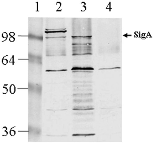
Background: We have previously shown that the enterotoxin SigA which resides on the she pathogenicity island (PAI) of S. flexneri 2a is an autonomously secreted serine protease capable of degrading casein. We have also demonstrated that SigA is cytopathic for HEp-2 cells and plays a role in the intestinal fluid accumulation associated with S. flexneri infections.

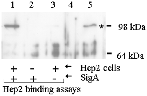
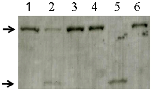
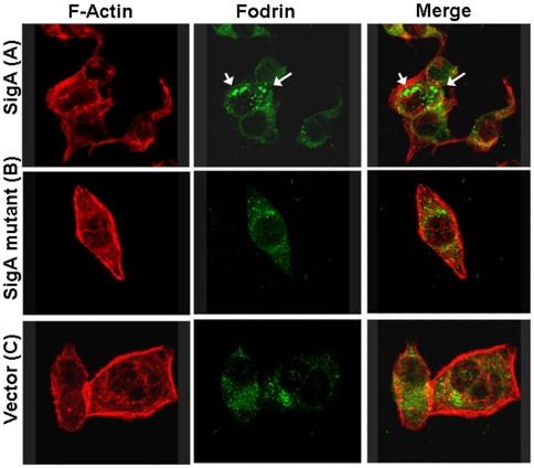
Methods/principal findings: In this work we show that SigA binds specifically to HEp-2 cells and degrades recombinant human alphaII spectrin (alpha-fodrin) in vitro, suggesting that the cytotoxic and enterotoxic effects mediated by SigA are likely associated with the degradation of epithelial fodrin. Consistent with our data, this study also demonstrates that SigA cleaves intracellular fodrin in situ, causing its redistribution within cells. These results strongly implicate SigA in altering the cytoskeleton during the pathogenesis of shigellosis. On the basis of these findings, cleavage of fodrin is a novel mechanism of cellular intoxication for a Shigella toxin. Furthermore, information regarding immunogenicity to SigA in infected patients is lacking. We studied the immune response of SigA from day 28 post-challenge serum of one volunteer from S. flexneri 2a challenge studies. Our results demonstrate that SigA is immunogenic following infection with S. flexneri 2a.

Conclusions: This work shows that SigA binds to epithelial HEp-2 cells as well as being able to induce fodrin degradation in vitro and in situ, further extending its documented role in the pathogenesis of Shigella infections.


Conflict of interest statement

Competing Interests: The authors have declared that no competing interests exist.

Figures

Figure 1
Figure 1. SigA binds specifically to HEp-2 cells.
HEp-2 cells were incubated in the presence (lane 1) or absence (lane 3) of SigA and washed to remove unbound SigA. Bound SigA was recovered by treatment of HEp-2 cells with protein sample buffer and assayed by Western analysis with anti-SigA antiserum. A control for non-specific binding of SigA to the tissue culture plate was also included (lane 2). Culture supernatants of E. coli either not expressing (lane 4) or expressing (lane 5) SigA, indicate the position of SigA on the gel (asterisk). The positions of standard molecular mass markers (kDa) are shown on the right.
Figure 2
Figure 2. Degradation of human α-fodrin by SigA.
Purified GST-fodrin was incubated with SigA (lanes 1, 2 and 3) or Pet positive control (lanes 4, 5 and 6) for zero (lanes 1 and 4) or 6 h (lanes 2 and 5). Lanes 3 and 6 shows SigA or Pet, respectively, pre-treated with PMSF and incubated with GST-fodrin for 6 h. The top arrow indicates the 109 kDa GST-fodrin fusion protein and the 74 kDa subproduct of degradation is indicated by the bottom arrow.
Figure 3
Figure 3. Effects of SigA on fodrin redistribution in epithelial cells.
HEp-2 cells were treated for 3 h with supernatant from SigA clone (A), or ΔSigA (B) or vector only used as a control (C). Cells were then fixed and stained with rhodamine-phalloidin, and anti-α-fodrin antibodies, and a fluorescein-labeled secondary anti-goat antibody. Intracellular aggregates of fodrin are indicated by arrows.
Figure 4
Figure 4. Antibody response against S. flexneri 2a SigA in a human volunteer.
Concentrated culture supernatant extracts were separated by SDS-PAGE and immunoreacted with human post-challenge serum. Lanes: 1, molecular mass markers (kDa); 2, supernatant of S. flexneri 2a YSH6000T (parent strain); 3, supernatant of SBA1356 (sepA and she deficient double mutant); 4, supernatant of SBA1359 (sigA-sepA-she triple mutant). The arrowhead indicates the reactivity of serum from a volunteer against the 103-kDa secreted SigA antigen.

References

    1. Kotloff KL, Winickoff JP, Ivanoff B, Clemens JD, Swerdlow DL, et al. Global burden of Shigella infections: implications for vaccine development and implementation of control strategies. Bull Wld Health Org. 1999;77:651–666. - PMC - PubMed
    1. Sasakawa, C Molecular basis of pathogenicity of Shigella. Rev Med Microbiol. 1995;6:257–266.
    1. Suzuki T, Sasakawa C. Molecular basis of the intracellular spreading of Shigella. Infect Immun. 2001;69:5959–5966. - PMC - PubMed
    1. Lan R, Lumb B, Ryan D, Reeves PR. Molecular evolution of large virulence plasmid in Shigella clones and enteroinvasive Escherichia coli. Infect Immun. 2001;69:6303–6309. - PMC - PubMed
    1. Henderson IR, Navarro-Garcia F, Nataro JP. The great escape: structure and function of the autotransporter proteins. Trends Microbiol. 1998;6:370–378. - PubMed

Publication types