Review
. 2010:2010:568068.
doi: 10.1155/2010/568068.

The mysterious unfoldome: structureless, underappreciated, yet vital part of any given proteome

Affiliations
Review

The mysterious unfoldome: structureless, underappreciated, yet vital part of any given proteome

Vladimir N Uversky. J Biomed Biotechnol. 2010.

Abstract

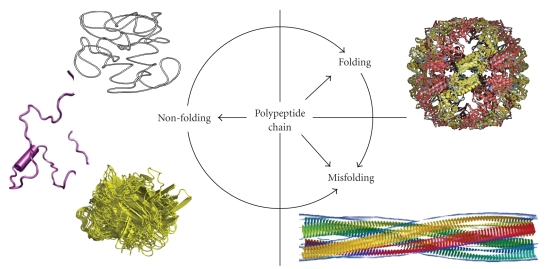
Contrarily to the general believe, many biologically active proteins lack stable tertiary and/or secondary structure under physiological conditions in vitro. These intrinsically disordered proteins (IDPs) are highly abundant in nature and many of them are associated with various human diseases. The functional repertoire of IDPs complements the functions of ordered proteins. Since IDPs constitute a significant portion of any given proteome, they can be combined in an unfoldome; which is a portion of the proteome including all IDPs (also known as natively unfolded proteins, therefore, unfoldome), and describing their functions, structures, interactions, evolution, and so forth. Amino acid sequence and compositions of IDPs are very different from those of ordered proteins, making possible reliable identification of IDPs at the proteome level by various computational means. Furthermore, IDPs possess a number of unique structural properties and are characterized by a peculiar conformational behavior, including their high stability against low pH and high temperature and their structural indifference toward the unfolding by strong denaturants. These peculiarities were shown to be useful for elaboration of the experimental techniques for the large-scale identification of IDPs in various organisms. Some of the computational and experimental tools for the unfoldome discovery are discussed in this review.


Figures

Figure 1
Figure 1
Fate of a polypeptide chain. Left. Three structures representing typical IDPs with different disorderedness levels (from top to bottom): native coil, native premolten globule, and native molten globule. Right. Top structure illustrates a well-folded protein, whereas the bottom structure represents one of the products of protein misfolding—a molecular model of the compact, 4-protofilament insulin fibril (http://people.cryst.bbk.ac.uk/~ubcg16z/amyloid/insmod.jpg).
Figure 2
Figure 2
Peculiarities of amino acid composition of ID proteins. (a) Comparison of the mean net charge and the mean hydrophobicity for a set of 275 ordered (open circles) and 91 natively unfolded proteins (gray circles). The solid line represents the border between extended IDPs and ordered proteins (see text). (b) Order/disorder composition profile. Comparisons of amino acid compositions of ordered proteins with each of three databases of disordered proteins. The ordinates are (%amino acid in disordered dataset - %amino acid in ordered dataset)/(%amino acid in ordered dataset) = ∆/globular_3D. Names of each database indicate how the disordered regions were identified. Negative values indicate that the disordered database has less than order, positive indicates more than order.
Figure 3
Figure 3
CH-CDF analysis of the genome-linked proteins VPgs from various viruses. Comparison of the results of PONDR CDF and CH-plot analyses for whole protein order-disorder via distributions of VPgs within the CH-CDF phase space. The protein analyzed by this approach are VPgs from Sobemovirus (Rice yellow mottle virus (RYMV), Cocksfoot mottle virus (CoMV), Ryegrass mottle virus (RGMoV), Southern bean mosaic virus (SBMV), Southern cowpea mosaic virus (SCPMV), Sesbania mottle virus (SeMV)), Potyvirus (Lettuce mosaic virus (LMV), Potato virus Y (PVY), Potato virus A (PVA), Tobacco etch virus (TEV), Turnip mosaic virus (TuMV), Bean yellow mosaic virus (BYMV)), and Caliciviridae (Rabbit hemorrhabic disease virus (RHDV), Vesicular exanthema of swine virus (VESV), Man Sapporo virus Manchester virus (SV), and Norwalk virus (NV)).
Figure 4
Figure 4
Schematic representation of the native/8 M urea 2D electrophoresis for separation of extended IDPs and globular proteins. A continuous line marks the diagonal of the gel to where IDPs run. A dashed line marks the position of globular proteins. A few proteins with a mixture of ordered and disordered regions are also indicated as “Partial IDPs”.

References

    1. Fischer E. Einfluss der configuration auf die wirkung der enzyme. Berichte der Deutschen Chemischen Gesellschaft. 1894;27(3):2985–2993.
    1. Lemieux RU, Spohr U. How Emil Fischer was led to the lock and key concept for enzyme specificity. Advances in Carbohydrate Chemistry and Biochemistry. 1994;50:1–20. - PubMed
    1. Dobson CM. Protein misfolding, evolution and disease. Trends in Biochemical Sciences. 1999;24(9):329–332. - PubMed
    1. Dunker AK, Oldfield CJ, Meng J, et al. The unfoldomics decade: an update on intrinsically disordered proteins. BMC Genomics. 2008;9(supplement 2, article S1) - PMC - PubMed
    1. Dunker AK, Brown CJ, Lawson JD, Iakoucheva LM, Obradović Z. Intrinsic disorder and protein function. Biochemistry. 2002;41(21):6573–6582. - PubMed

Publication types

LinkOut - more resources