

Renal fibrosis
Abstract
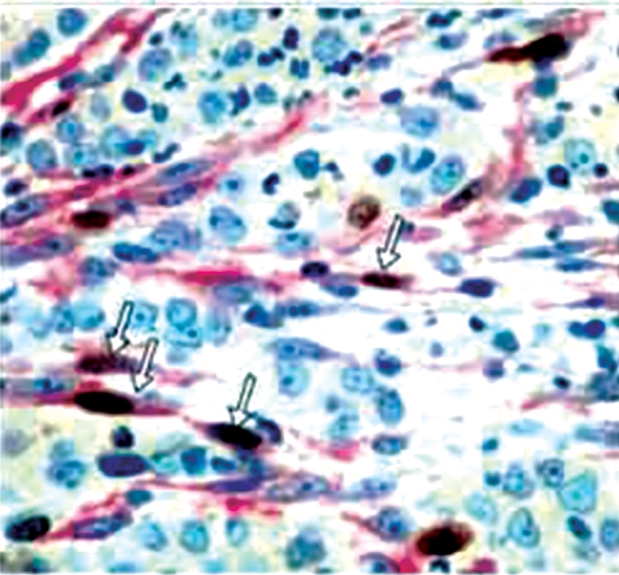
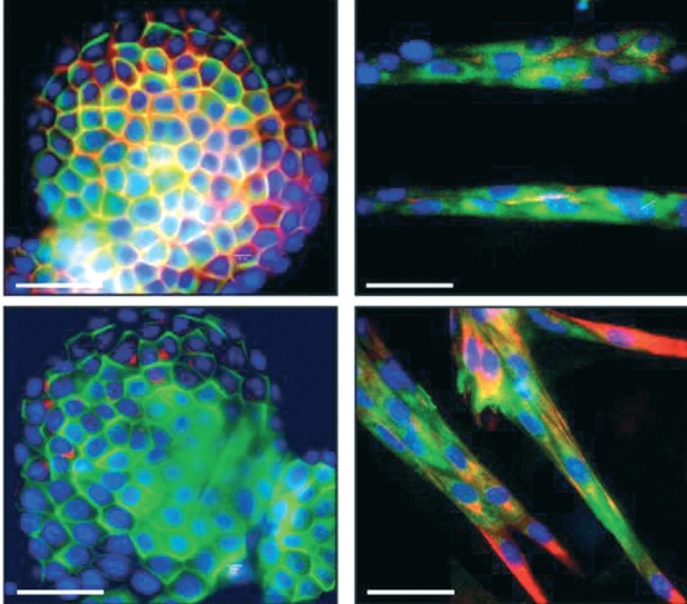
Tubulointerstitial renal fibrosis, characterized as a progressive detrimental connective tissue deposition on the kidney parenchyma, appears to be a harmful process leading inevitably to renal function deterioration, independently of the primary renal disease which causes the original kidney injury. Epithelial to Mesenchymal Transition (EMT) of tubular epithelial cells which are transformed to mesenchymal fibroblasts migrating to adjacent interstitial parenchyma constitutes the principal mechanism of renal fibrosis along with local and circulating cells. Proteinuria as well as hypoxia is included among the main mechanisms of EMT stimulation. TGFbeta-1 through the SMAD pathway is considered as the main modulator regulating the EMT molecular mechanism, probably in cooperation with hypoxia inducible factors. Hepatocyte Growth Factor (HGF) and Bone Morphogenetic Factor-7 (BMF-7) are inhibitory to EMT molecules which could prevent in experimental and clinical level the catastrophic process of interstitial fibrosis. Interesting data emerge indicating that HGF and BMF-7 administration prevents the peritoneal fibrosis of mesothelial cells.
Keywords: TGF-β; angiotensin II; cytokines; epithelial mesenchymal transition; fibrosis; growth factors; hypoxia; proteinuria.
Figures
Similar articles
-
Novel RAS inhibitor 25-O-methylalisol F attenuates epithelial-to-mesenchymal transition and tubulo-interstitial fibrosis by selectively inhibiting TGF-β-mediated Smad3 phosphorylation.Phytomedicine. 2018 Mar 15;42:207-218. doi: 10.1016/j.phymed.2018.03.034. Epub 2018 Mar 19. Phytomedicine. 2018. PMID: 29655688
-
Lefty-1 inhibits renal epithelial-mesenchymal transition by antagonizing the TGF-β/Smad signaling pathway.J Mol Histol. 2020 Feb;51(1):77-87. doi: 10.1007/s10735-020-09859-8. Epub 2020 Feb 17. J Mol Histol. 2020. PMID: 32065356
-
FSP1-specific SMAD2 knockout in renal tubular, endothelial, and interstitial cells reduces fibrosis and epithelial-to-mesenchymal transition in murine STZ-induced diabetic nephropathy.Cell Tissue Res. 2018 Apr;372(1):115-133. doi: 10.1007/s00441-017-2754-1. Epub 2017 Dec 6. Cell Tissue Res. 2018. PMID: 29209813
-
Obstructive nephropathy and renal fibrosis: The role of bone morphogenic protein-7 and hepatocyte growth factor.Kidney Int Suppl. 2003 Nov;(87):S105-12. doi: 10.1046/j.1523-1755.64.s87.16.x. Kidney Int Suppl. 2003. PMID: 14531782 Review.
-
Pathogenesis of the podocytopathy and proteinuria in diabetic glomerulopathy.Curr Diabetes Rev. 2008 Feb;4(1):39-45. doi: 10.2174/157339908783502370. Curr Diabetes Rev. 2008. PMID: 18220694 Review.
Cited by
-
Treadmill Exercise Training Ameliorates Functional and Structural Age-Associated Kidney Changes in Male Albino Rats.ScientificWorldJournal. 2021 Nov 30;2021:1393372. doi: 10.1155/2021/1393372. eCollection 2021. ScientificWorldJournal. 2021. PMID: 34887703 Free PMC article.
-
The renal consequences of maternal obesity in offspring are overwhelmed by postnatal high fat diet.PLoS One. 2017 Feb 22;12(2):e0172644. doi: 10.1371/journal.pone.0172644. eCollection 2017. PLoS One. 2017. PMID: 28225809 Free PMC article.
-
The promising shadow of microbubble over medical sciences: from fighting wide scope of prevalence disease to cancer eradication.J Biomed Sci. 2021 Jun 21;28(1):49. doi: 10.1186/s12929-021-00744-4. J Biomed Sci. 2021. PMID: 34154581 Free PMC article. Review.
-
Acute Decompensated Heart Failure and the Kidney: Physiological, Histological and Transcriptomic Responses to Development and Recovery.J Am Heart Assoc. 2021 Sep 21;10(18):e021312. doi: 10.1161/JAHA.121.021312. Epub 2021 Sep 17. J Am Heart Assoc. 2021. PMID: 34533033 Free PMC article.
-
Targeting lysine-specific demethylase 1A inhibits renal epithelial-mesenchymal transition and attenuates renal fibrosis.FASEB J. 2022 Jan;36(1):e22122. doi: 10.1096/fj.202101566R. FASEB J. 2022. PMID: 34958158 Free PMC article.
References
-
- Robertson H, Kirby JA, Yip WW, Jones DE, Burt AD. Biliary epithelial-mesenchymal transition in posttransplantation reccurence of primary biliary cirrhosis. Hepatology. 2007;45:977–981. - PubMed
-
- Willis BC, Boroc Z. TGF-beta-induced EMT: mechanisms and implications for fibrotic lung disease. Am J Physiol Lung Cell Mol Physiol. 2007;293:L525–L534. - PubMed
-
- Liu Y. Epithelial to Mesenchymal Transition in Renal Fibrogenesis: Pathologic Significance, Molecular Mechanism, and Therapeutic Intervention. J Am Soc Nephrol. 2004;15:1–12. - PubMed
LinkOut - more resources
Full Text Sources