. 2009 Dec;5(12):e1000687.
doi: 10.1371/journal.ppat.1000687. Epub 2009 Dec 11.

CD14 signaling restrains chronic inflammation through induction of p38-MAPK/SOCS-dependent tolerance

Affiliations

CD14 signaling restrains chronic inflammation through induction of p38-MAPK/SOCS-dependent tolerance

Bikash Sahay et al. PLoS Pathog. 2009 Dec.

Abstract

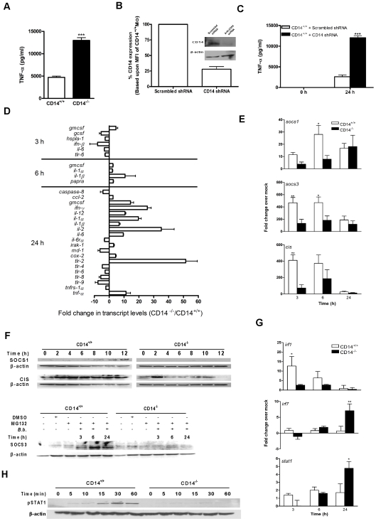
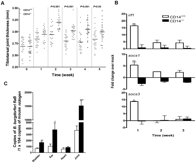
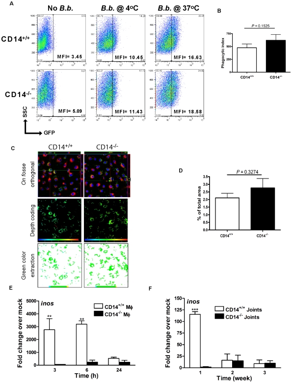
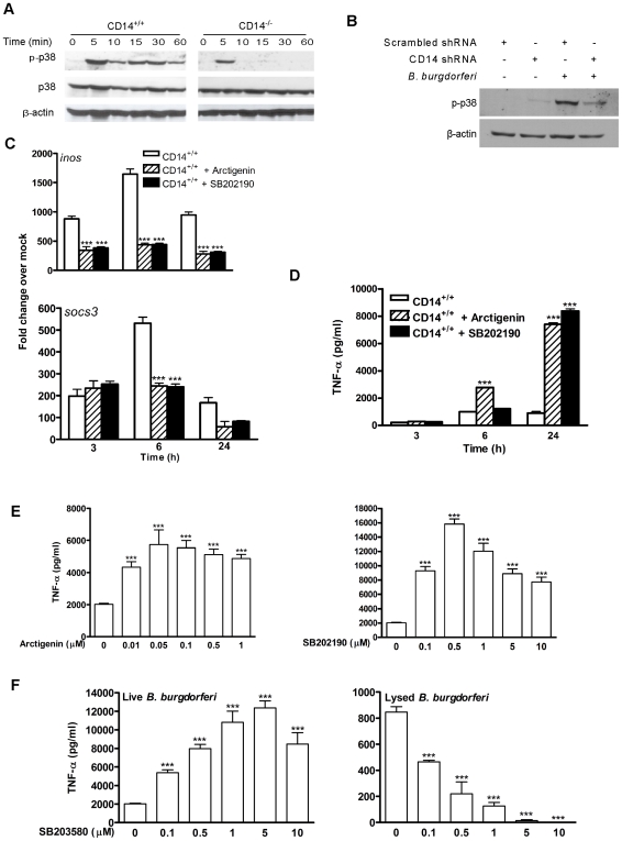
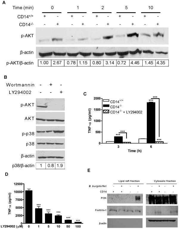
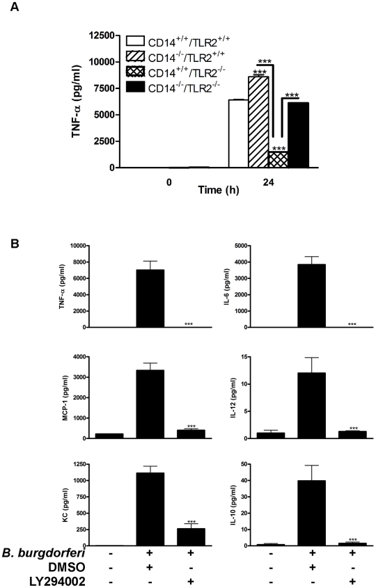
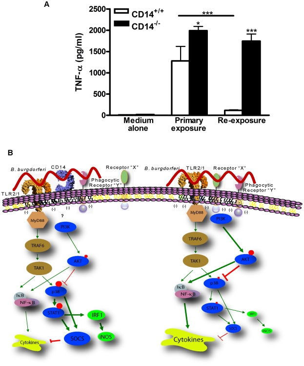
Current thinking emphasizes the primacy of CD14 in facilitating recognition of microbes by certain TLRs to initiate pro-inflammatory signaling events and the importance of p38-MAPK in augmenting such responses. Herein, this paradigm is challenged by demonstrating that recognition of live Borrelia burgdorferi not only triggers an inflammatory response in the absence of CD14, but one that is, in part, a consequence of altered PI3K/AKT/p38-MAPK signaling and impaired negative regulation of TLR2. CD14 deficiency results in increased localization of PI3K to lipid rafts, hyperphosphorylation of AKT, and reduced activation of p38. Such aberrant signaling leads to decreased negative regulation by SOCS1, SOCS3, and CIS, thereby compromising the induction of tolerance in macrophages and engendering more severe and persistent inflammatory responses to B. burgdorferi. Importantly, these altered signaling events and the higher cytokine production observed can be mimicked through shRNA and pharmacological inhibition of p38 activity in CD14-expressing macrophages. Perturbation of this CD14/p38-MAPK-dependent immune regulation may underlie development of infectious chronic inflammatory syndromes.


Conflict of interest statement

The authors have declared that no competing interests exist.

Figures

Figure 1
Figure 1. Higher B. burgdorferi-induced inflammatory gene activity is associated with lower SOCS levels in CD14−/− MΦ.
A) MΦ isolated from CD14+/+ and CD14−/− mice were incubated with B. burgdorferi at a MOI of 10 for 24 h and TNF-α levels in culture supernatant were measured by CBA. B) Lentiviral transduction was used to knock down CD14 in MΦ as determined by a reduction in mean fluorescent intensity (MFI) and by Western blot analysis (inset). C) The lentivirus-treated MΦ were incubated with B. burgdorferi for 24 h and TNF-α levels were measured as for (A). D) MΦ isolated from CD14+/+ and CD14−/− mice were incubated with B. burgdorferi for 3, 6 and 24 h. Total RNA was isolated and used to perform qPCR for simultaneous interrogation of 84 genes associated with TLR signaling. The results presented are the ratio of fold change in HPRT-normalized gene activity in CD14−/− versus CD14+/+ MΦ and error bars represent SEM calculated on the basis of three independent experiments. E) Total RNA isolated from CD14+/+ and CD14−/− MΦ incubated with B. burgdorferi was analyzed by qPCR for socs1, socs3 and cis. Results are presented as fold change over respective mock-infected control and were normalized with 18S rRNA transcript. F) Equivalent protein from lysed MΦ were separated by 12% SDS-PAGE, transferred to a PVDF membrane and probed with antibodies directed against SOCS1, SOCS3, CIS or β-actin. G) Total RNA isolated from CD14+/+ and CD14−/− MΦ incubated with B. burgdorferi was analyzed by qPCR for irf1, irf7 and stat1. Dotted lines denote the 2-fold over mock change considered significant for the purposes of interpretation. H) Equivalent protein from lysed MΦ were subjected to Western blot analysis using antibodies directed against phospho-specific STAT1 or STAT3, or β-actin. Results represent mean±SEM from three to four independent experiments. *P<0.05, **P<0.01, ***P<0.001.
Figure 2
Figure 2. CD14 deficiency is associated with greater inflammation and higher bacterial burden with reduced socs transcription in response to B. burgdorferi.
A) CD14+/+ and CD14−/− mice were infected with 1×10⁵ B. burgdorferi and tibiotarsal joint thickness was measured at 1 week intervals. The horizontal bars indicate mean thickness for each group and the data are representative of two independent experiments (n = 24). B) Total RNA was isolated from the tibiotarsal joints of CD14+/+ and CD14−/− mice (n = 6), an equal amount of RNA was pooled from each joint and 0.5 µg of pooled RNA was used for preparing cDNA. The cDNA was analyzed by qPCR to determine the levels of irf1, socs1 and socs3 transcript. Results represent mean±SEM from two independent experiments wherein samples were run in triplicate. *P<0.05, **P<0.01. C) DNA from the indicated organs was collected 6 weeks p.i. and the bacterial burden was determined using Taqman® probes for flaB of B. burgdorferi; murine nidogen served as a control. Results represent mean±SEM from two independent experiments (n = 24) wherein samples were run in triplicate. *P<0.05, **P<0.01.
Figure 3
Figure 3. B. burgdorferi-induced transcription of inos, but not phagocytic uptake of spirochetes, is impaired by CD14 deficiency.
A) MΦ were incubated with GFP-expressing B. burgdorferi at a MOI of 10 at 4°C and 37°C for 6 h. Phagocytosis of B. burgdorferi was determined by flow cytometry. B) Phagocytic indices were calculated based upon the results of five independent flow cytometric experiments. C) MΦ were incubated with GFP-expressing B. burgdorferi at a MOI of 100 for 6 h and the cytoplasm and nucleus were stained with wheat-germ agglutinin-Alexa Fluor 647 and DAPI, respectively. Optical sections were collected at every 0.5µm and were used to generate orthogonal sections (upper panel), depth coded images (middle panel) and green color extraction (lower panel) (see Methods). D) The green area of 10 randomly chosen fields from both genotypes was used for statistical analysis. E) inos transcript levels were determined by qPCR using RNA isolated from MΦ incubated with B. burgdorferi or F) using RNA pooled from joints isolated from infected mice, as described in Figure 2. Results represent mean±SEM from two to five independent experiments. **P<0.01, ***P<0.001.
Figure 4
Figure 4. CD14 deficiency results in dysregulated p38-MAPK signaling and cytokine production in response to B. burgdorferi.
A) Equal protein from lysates of CD14+/+ and CD14−/− MΦ incubated with B. burgdorferi were separated by 10% SDS-PAGE, transferred to a nitrocellulose membrane and probed with phospho-p38 and β-actin antibodies. B) CD14 was knocked down using lentiviral transduction as in Figure 1B & C. MΦ were incubated with B. burgdorferi for 30 min and phospho-p38 and β-actin were detected as in Figure 4A. C) CD14+/+ MΦ were treated with DMSO, SB202190 (0.5 µM) or arctigenin (1 µM) for 30 min prior to incubation with B. burgdorferi and total RNA was analyzed by qPCR for inos and socs3. D) CD14+/+ MΦ were treated as described in (C), and culture supernatants were analyzed for TNF-α by CBA. E) CD14+/+ MΦ were treated with DMSO or increasing concentrations of arctigenin or SB202190 for 30 min prior to incubation with B. burgdorferi. Cell culture supernatants were collected 24 h p.i. and TNF-α was measured by CBA. F) CD14+/+ MΦ were treated with DMSO or increasing concentrations of SB203580 for 30 min prior to incubation with live B. burgdorferi or B. burgdorferi lysate (10µg/ml). Cell culture supernatants were assayed for TNF-α by CBA. Results represent mean±SEM from two to five independent experiments. *P<0.05, **P<0.01, ***P<0.001.
Figure 5
Figure 5. Inhibition of B. burgdorferi-induced AKT activation in CD14−/− MΦ reestablishes p38 activity and restores negative regulation of cytokine production.
A) Equal protein from lysed CD14+/+ and CD14−/− MΦ incubated with B. burgdorferi were separated by SDS-PAGE, transferred to a nitrocellulose membrane and probed for phospho-AKT and β-actin. B) CD14−/− MΦ were treated with DMSO or PI3K inhibitors [wortmannin (100nM) or Ly294002 (100µM)] for 30 min prior to incubation with B. burgdorferi. Western blots were probed with phospho-specific AKT and p38 antibodies, then striped and reprobed for total AKT, p38 and β-actin. C) CD14−/− MΦ were treated with Ly294002 (100µM) for 30 min prior to incubation with B. burgdorferi for 3 and 6 h. Culture supernatants were assayed for TNF-α by CBA. TNF-α release by stimulated CD14+/+ MΦ served as a control. D) CD14−/− MΦ were treated with DMSO or LY294002 for 30 min prior to incubation with B. burgdorferi. Culture supernatants were collected 24 h p.i. and TNF-α was measured by CBA. E) Lipid rafts were isolated from CD14+/+ and CD14−/− MΦ incubated with or without B. burgdorferi at a MOI of 10 for 5 min. An equal volume of lipid raft and cytosolic fractions were separated by 10% SDS-PAGE, transferred to a nitrocellulose membrane and probed with antibodies directed against PI3K, flotillin-1, and β-actin. Results represent mean±SEM from two to five independent experiments. ***P<0.001.
Figure 6
Figure 6. TLR2 plays a partial role in both CD14-dependent and -independent cytokine production.
A) MΦ isolated from CD14+/+, CD14−/−, TLR2−/− and CD14−/−/TLR2−/− mice were incubated with B. burgdorferi and culture supernatants were assayed for TNF-α by CBA. B) B. burgdorferi was incubated with MΦ isolated from CD14−/−/TLR2−/− mice in the presence of DMSO or 100µM Ly294002 for 24 h and cytokines were measured by CBA. Results represent mean±SEM from three independent experiments. ***P<0.001.
Figure 7
Figure 7. CD14-dependent signaling is a requirement for B. burgdorferi-induced tolerance in MΦ.
A) CD14+/+ and CD14−/− MΦ were incubated with B. burgdorferi at a MOI of 10 for 12 h (primary exposure). Cell monolayers were washed extensively followed by reexposure (secondary exposure) to medium containing B. burgdorferi for an additional 6 h. The TNF-α level in culture supernatants from cells rexposed to spirochetes was compared with levels released by MΦ which received only a primary exposure to B. burgdorferi. B) A schematic diagram illustrating CD14-dependent and -independent signaling events in response to B. burgdorferi. In the absence of CD14 neither binding nor uptake of B. burgdorferi via a putative Phagocytic Receptor “Y” is affected. However, localization of PI3K to the signaling platform is altered, perhaps owing to the presence of Receptor “X”, which increases the pool of phospho-AKT resulting in only transient p38 activity. Impaired p38 activity correlates with increased transcription and expression of TLR2, reduced activation of STAT1 and IRF1, and decreased induction of SOCS, all of which are necessary to modulate the intensity and duration of Lyme borreliosis. Ultimately, it is CD14-dependent induction of p38-mediated SOCS activity which regulates the intensity and duration of macrophage response to B. burgdorferi through the induction of tolerance. The size of the red circles indicates the degree of phosphorylation of the indicated molecule and the size of the individual molecules is reflective of relative transcript/protein levels. The thickness of lines in the model reflects the intensity of the positive or negative action on downstream molecules.

Similar articles

Cited by

References

    1. Wooten RM, Weis JJ. Host-pathogen interactions promoting inflammatory Lyme arthritis: use of mouse models for dissection of disease processes. Curr Opin Microbiol. 2001;4:274–279. - PubMed
    1. Arbibe L, Mira JP, Teusch N, Kline L, Guha M, et al. Toll-like receptor 2-mediated NF-[kappa]B activation requires a Rac1-dependent pathway. Nat Immunol. 2000;1:533–540. - PubMed
    1. Steere AC, Glickstein L. Elucidation of Lyme arthritis. Nat Rev Immunol. 2004;4:143–152. - PubMed
    1. Antal S. Evaluation of CD14 in host defence. European Journal of Clinical Investigation. 2000;30:167–179. - PubMed
    1. Sellati TJ, Bouis DA, Kitchens RL, Darveau RP, Pugin J, et al. Treponema pallidum and Borrelia burgdorferi lipoproteins and synthetic lipopeptides activate monocytic cells via a CD14-dependent pathway distinct from that used by lipopolysaccharide. J Immunol. 1998;160:5455–5464. - PubMed

Publication types

MeSH terms

Substances