. 2009 Dec;5(12):e1000690.
doi: 10.1371/journal.ppat.1000690. Epub 2009 Dec 11.

Functional memory B cells and long-lived plasma cells are generated after a single Plasmodium chabaudi infection in mice

Affiliations

Functional memory B cells and long-lived plasma cells are generated after a single Plasmodium chabaudi infection in mice

Francis Maina Ndungu et al. PLoS Pathog. 2009 Dec.

Abstract

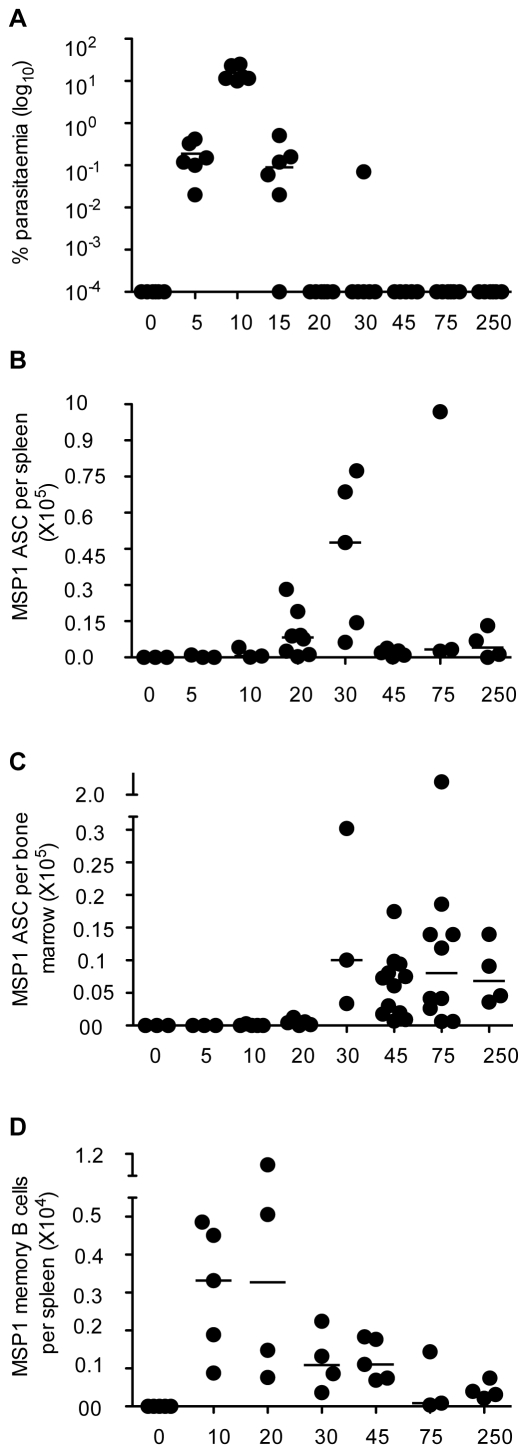
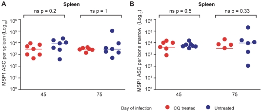
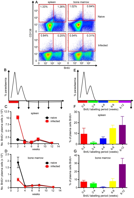
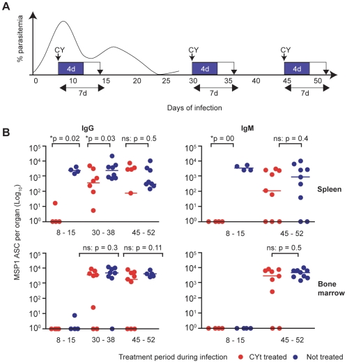
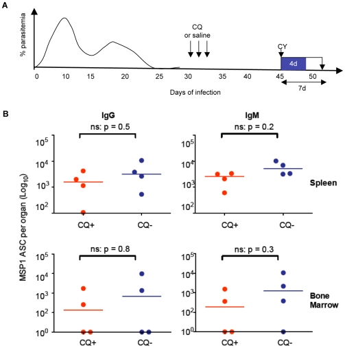
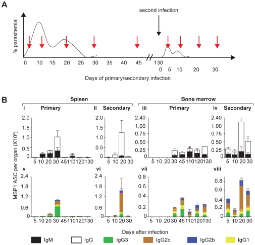
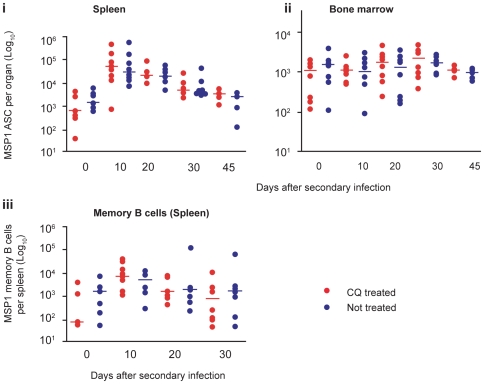
Antibodies have long been shown to play a critical role in naturally acquired immunity to malaria, but it has been suggested that Plasmodium-specific antibodies in humans may not be long lived. The cellular mechanisms underlying B cell and antibody responses are difficult to study in human infections; therefore, we have investigated the kinetics, duration and characteristics of the Plasmodium-specific memory B cell response in an infection of P. chabaudi in mice. Memory B cells and plasma cells specific for the C-terminal region of Merozoite Surface Protein 1 were detectable for more than eight months following primary infection. Furthermore, a classical memory response comprised predominantly of the T-cell dependent isotypes IgG2c, IgG2b and IgG1 was elicited upon rechallenge with the homologous parasite, confirming the generation of functional memory B cells. Using cyclophosphamide treatment to discriminate between long-lived and short-lived plasma cells, we demonstrated long-lived cells secreting Plasmodium-specific IgG in both bone marrow and in spleens of infected mice. The presence of these long-lived cells was independent of the presence of chronic infection, as removal of parasites with anti-malarial drugs had no impact on their numbers. Thus, in this model of malaria, both functional Plasmodium-specific memory B cells and long-lived plasma cells can be generated, suggesting that defects in generating these cell populations may not be the reason for generating short-lived antibody responses.


Conflict of interest statement

The authors have declared that no competing interests exist.

Figures

Figure 1
Figure 1. Kinetics of P. chabaudi parasitemia and the contemporaneous B cell responses following a primary infection.
(A) A representative experiment showing the percentage of P. chabaudi iRBC after infection of C57BL/6 mice with 105 P. chabaudi iRBC. Each symbol represents an individual mouse (n = 6) and the horizontal bars show the median values for each time point. (All values of Y = 10−4 or less are overlaid on the X-axis and appear as a single symbol). After day 30 of infection, the percentage parasitaemias were generally either 10−4 (day 45, 5/6 mice) or below detection (day 75). However, presence of parasites can be demonstrated by transferring infected blood into immunocompromised mice for up to 3 months . (B–D) The numbers of MSP-1 specific ASC in spleen and bone marrow, and the number of memory B cells (MBC) were determined at different time points of the P. chabaudi infection by ex vivo (ASC) and in vitro cultured (MBC) ELISpot assays as described in the experimental procedures. (B) Total number of MSP119-specific IgG Ab secreting cells (ASC) in the spleens at different times after infection. (C) Total number of MSP119-specific IgG ASC in bone marrow at different times of a P. chabaudi infection, calculated as described in the experimental procedures. (D) Total numbers of MSP119-specific IgG MBC in spleens of infected and control spleens after polyclonal stimulation and differentiation into Ab secreting cells in in vitro limiting dilution cultures. For (B, C and D) each symbol represents the number of ELISpots in the organs of an individual mouse after subtraction of the mean background value of 3 naïve control mice. Each time point shows ELISpot values from 3 to 8 mice, and the horizontal bars represent the median values. All values of Y = 0 or less are overlaid on the X-axis and appear as single symbols). Changes in numbers of ASC/MBC from one time point to the next were determined using a Mann Whitney test; where significant p values are shown in the text.
Figure 2
Figure 2. MSP119-specific IgG ASC are maintained independently of low-grade chronic parasitemias.
Total numbers of MSP119-specific IgG ASC in spleen (A) and bone marrow (B) were determined in chloroquine-treated (red symbols, n = 7) or untreated (blue symbols, n = 7) mice, 45 and 75 days after infection of C57BL/6 mice with 105 P. chabaudi iRBC. Chloroquine (25 mg/kg body weight) or saline was administered after resolution of the acute phase of parasitemia as described in the experimental procedures. Each dot represents the values obtained from a single mouse after subtraction of the background value of 3 uninfected control mice as described in Figure 1. The horizontal lines indicate the medians for each group at each time point. Differences between ASC in the 2 groups at each time point were determined by a Mann Whitney test; the p values are shown and ns indicates no significant differences.
Figure 3
Figure 3. Plasma cells generated in the first 2 weeks of an acute P. chabaudi infection are not long-lived.
(A) Cartoon indicating the 2-week period of oral BrdU administration after infection of C57BL/6 mice with 105 P. chabaudi iRBC, and the subsequent timing of removal of spleens and bone marrow for the analysis shown in graphs B & C. Total numbers of BrdU-labelled CD138+ cells (gated as shown in Supplementary Figure S3) in spleen (B) and bone marrow (C) of P. chabaudi-infected mice. (D) Cartoon indicating the different 2- or 4-week time periods of oral BrdU administration following infection of C57BL/6 mice with 105 P. chabaudi iRBC for the analysis for graphs E & F. Spleens and bone marrows were removed and analysed after 12 weeks of infection. Percentage of CD138+ cells labelled with BrdU (gated as shown in Supplementary Figure S3) in spleens (E) and bone marrow (F) of infected mice at 12 weeks post-infection. The values shown are the mean number (B and C) or percentage of cells (E and F) from 5 individual mice, and the error bars represent the standard errors of the means.
Figure 4
Figure 4. Intrinsically long-lived MSP119-specific IgG and IgM ASC are generated in a primary infection.
(A) Cartoon showing the time course of the experiment. C57BL/6 mice were infected with 105 P. chabaudi iRBC, and received either 140 mg Cyclophosphamide (CY) per Kg body weight (over 4 days), or normal saline (control) at days 8, 30 and 45. Sampling of spleens and bone marrows were carried out 7 days later at days 15, 38 and 52 days. The numbers of splenic and bone-marrow IgG ASC were determined by ex vivo ELISpot assays as described in the experimental procedures. (B) Left panel, Comparison of numbers of MSP119-specific IgG ASC between control treated mice (blue circles) and CY treated mice (red circles) at days 8–15 (n = 4), 30–38 (n = 8) and 45–52 (n = 9). Right panel, Comparison of numbers of MSP119-specific IgM ASC between CY treated and control mice at 8–15 (n = 4) and 45–52 (n = 9) time intervals of treatment and sampling. (No MSP119-specific ASC were detected in the bone marrow at day 15). Background values from naive uninfected mice have been subtracted from all the values shown as described in Figure 1. Each symbol represents an individual mouse, and the horizontal bars indicate the median values of 8 mice. The numbers of ASC in the two groups at each time point were compared using a Mann Whitney test; * indicates the differences were significant (p values are shown), and ns no significant differences.
Figure 5
Figure 5. Low-grade P. chabaudi chronic infection does not affect ASC longevity.
(A) Cartoon showing the course of the experiment. To test whether low-grade chronic infections of P. chabaudi in mice affect the longevity of MSP119-specific ASC, C57Bl/6 mice infected with 105 P. chabaudi iRBC were either given the antimalarial drug CQ or saline (indicated by arrows) as described in the experimental procedures. Both groups of mice were then treated with 140mg CY/Kg body weight at day 45 of infection as described in the experimental procedures (indicated by the arrow and blue box) and sacrificed 7 days later for the determination of numbers of MSP119-specific ASC. (B) Comparison of the total numbers of MSP119-specific IgG ASC (left panel) and MSP119-specific IgM ASC (right) panel in the spleens (upper graphs) and bone marrow (lower graphs) of CQ-treated (red circles) and untreated control (blue circles) mice. Background values from naive uninfected mice have been subtracted from all the values shown. Each symbol represents an individual mouse, and the horizontal bars indicate the median values of 4 mice. The naïve background was zero for each of the four graphs. The numbers of ASC in the two groups at each time point were compared using a Mann Whitney test; * indicates the differences were significant (p values are shown), and ns no significant differences.
Figure 6
Figure 6. Evidence for generation and maintenance of functional memory B cell in a primary infection.
(A) Cartoon showing the course of the experiment. To compare the kinetics and distribution of ASC secreting different isotypes of anti-MSP119 Ab between primary and secondary infections, C57Bl/6 mice were infected with 105 P. chabaudi iRBC and then rested for 130 days. They were then re-infected with the same dose of iRBC (black arrow), together with previously naïve age-matched controls (primary infection). Mice from both groups (primary and secondary infections) were then sacrificed at the different time points (red arrows) and numbers of MSP119-specific ASCs of different isotypes present in spleens and bone marrow determined in ex vivo ELISpot assays. (B) (i, ii) Relative numbers of MSP119-specific IgG (open bars) and IgM (filled bars) specific ASC in spleens of infected mice following primary and secondary infections at different time points following infection. (iii, iv) Relative numbers of MSP119-specific IgG (open bars) and IgM (filled bars) specific ASC in bone marrow of infected mice following primary and secondary infections at different time points following infection. (v, vi) Relative numbers of MSP119-specific IgG subclass ASC (IgG1 [yellow bars], IgG2c [brown bars], IgG2b [blue bars] and IgG3 [green bars]) specific ASC in spleens of infected mice following primary and secondary infections at different time points following infection. (vii, viii) Relative numbers of MSP119-specific IgG subclass ASC (IgG1 [yellow bars], IgG2c [brown bars], IgG2b [blue bars] and IgG3 [green bars]) specific PC in bone marrow of infected mice following primary and secondary infections at different time points following infection. In each case, the means and standard errors of the mean for 3 to 5 mice at each time point are shown.
Figure 7
Figure 7. Persistent low-grade chronic P. chabaudi infection does not affect MSP119-specific memory B cell responses.
(A) Cartoon showing the course of the experiment. Infected mice were treated either with 25 mg/Kg body weight of chloroquine (CQ) from day 30 of infection as described in Figure 2, or with normal saline (controls), and the number of MSP119 IgG ASC present after a second infection were compared between the two groups. (B) (i) Comparison of numbers of MSP119-specific IgG ASC in the spleens of CQ-treated (red circles) and untreated mice (blue circles). (ii) Comparison of numbers of MSP119-specific IgG ASC in the bone marrow of treated (red circles) and untreated (blue circles). (iii) Comparison of numbers of MSP119-specific IgG MBC in the spleens of treated (red dots) and untreated (blue dots). In each case, medians of 5 to 8 individual mice are shown. The numbers of ASC in the two groups at each time point were compared using a Mann Whitney test; * indicates the differences were significant (p values are shown), and ns no significant differences.

Similar articles

Cited by

References

    1. Cavanagh DR, Dodoo D, Hviid L, Kurtzhals JA, Theander TG, et al. Antibodies to the N-terminal block 2 of Plasmodium falciparum merozoite surface protein 1 are associated with protection against clinical malaria. Infect Immun. 2004;72:6492–6502. - PMC - PubMed
    1. Cohen S, McGregor IA, Carrington S. Gamma-globulin and acquired immunity to human malaria. Nature. 1961;192:733–737. - PubMed
    1. Conway DJ, Cavanagh DR, Tanabe K, Roper C, Mikes ZS, et al. A principal target of human immunity to malaria identified by molecular population genetic and immunological analyses. Nat Med. 2000;6:689–692. - PubMed
    1. Metzger WG, Okenu DM, Cavanagh DR, Robinson JV, Bojang KA, et al. Serum IgG3 to the Plasmodium falciparum merozoite surface protein 2 is strongly associated with a reduced prospective risk of malaria. Parasite Immunol. 2003;25:307–312. - PubMed
    1. Osier FH, Fegan G, Polley SD, Murungi L, Verra F, et al. Breadth and magnitude of antibody responses to multiple Plasmodium falciparum merozoite antigens are associated with protection from clinical malaria. Infect Immun. 2008;76:2240–2248. - PMC - PubMed

Publication types

Substances