. 2007 Aug;20(3):148-57.
doi: 10.1055/s-2007-984859.

Rectal cancer staging

Affiliations

Rectal cancer staging

James S Wu. Clin Colon Rectal Surg. 2007 Aug.

Abstract

Rectal cancer staging provides critical information concerning the extent of the disease. The information gained from staging is used to determine prognosis, to guide management, and to assess response to therapy. Accurate staging is essential for directing the multidisciplinary approach to therapy. This article focuses on the evolution of staging systems, the rational for staging, and current methods used to stage rectal cancer.

Keywords: Rectal cancer; computed tomography; endorectal ultrasound; magnetic resonance imaging; positron emission tomography; staging.


Figures
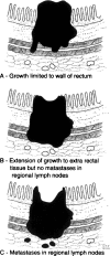
Figure 1
Figure 1
Rectal cancer staging system proposed by Dukes in 1932. (From the Cleveland Clinic Foundation, Cleveland, Ohio. Reprinted with permission.)
Figure 2
Figure 2
Diagrammatic representation of rectal thirds as defined by sigmoidoscopic distance from the anal canal (after Goligher et al16). (From the Cleveland Clinic Foundation, Cleveland, Ohio. Reprinted with permission.)
Figure 3
Figure 3
Five-layer model for the interpretation of endorectal ultrasound. Three echogenic (white lines) and two echo-poor (dark lines) are seen (after Orrom et al36). (From the Cleveland Clinic Foundation, Cleveland, Ohio. Reprinted with permission.)
Figure 4
Figure 4
Endorectal ultrasound of a uT3 rectal cancer. The outer black line is breached. The scalloped pattern of the outer tumor edge is consistent with penetration of the muscularis mucosa (large arrow). Note the five layers of the normal rectal wall (small arrow). (From the Cleveland Clinic Foundation, Cleveland, Ohio. Reprinted with permission.)
Figure 5
Figure 5
(A) Magnetic resonance imaging (MRI) scan of a rectal cancer showing the circumferential mesorectal envelope. (B) MRI scan of the rectal cancer patient depicted in (A) showing lymph nodes. (C) MRI scan showing locally advanced rectal cancer that has invaded the posterior wall of the vagina (arrow). (From the Cleveland Clinic Foundation, Cleveland, Ohio. Reprinted with permission.)

Similar articles

Cited by

References

    1. Jemal A, Siegel R, Ward E, Murray T, Xu J, Thun M J. Cancer statistics, 2007. CA Cancer J Clin. 2007;57:43–66. - PubMed
    1. Dukes C E. The classification of cancer of the rectum. J Pathol Bacteriol. 1932;35:323–332.
    1. Zinkin L D. A critical review of the classifications and staging of colorectal cancer. Dis Colon Rectum. 1983;26:37–43. - PubMed
    1. Lockhart-Mummery J P. Two hundred cases of cancer of the rectum treated by perineal excision. Br J Surg. 1926–1927;14:110–124.
    1. Kirklin J W, Dockerty M B, Waugh J M. The role of the peritoneal reflection in the prognosis of carcinoma of the rectum and sigmoid colon. Surg Gynecol Obstet. 1949;88:326–331. - PubMed