. 2009 Nov 27:3:26.
doi: 10.3389/neuro.05.026.2009. eCollection 2009.

Primate-specific origins and migration of cortical GABAergic neurons

Affiliations

Primate-specific origins and migration of cortical GABAergic neurons

Zdravko Petanjek et al. Front Neuroanat. .

Abstract

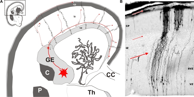
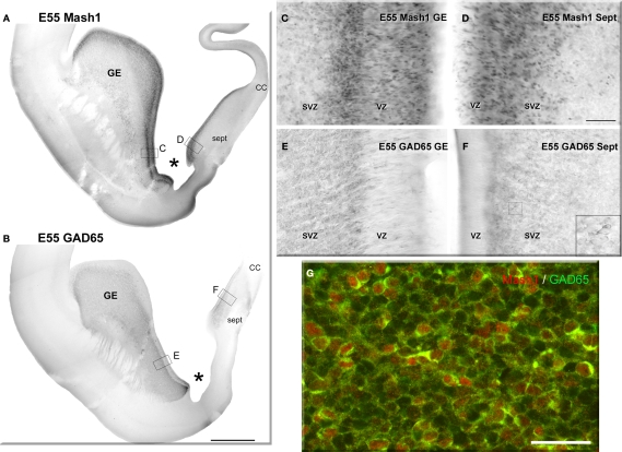
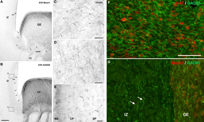
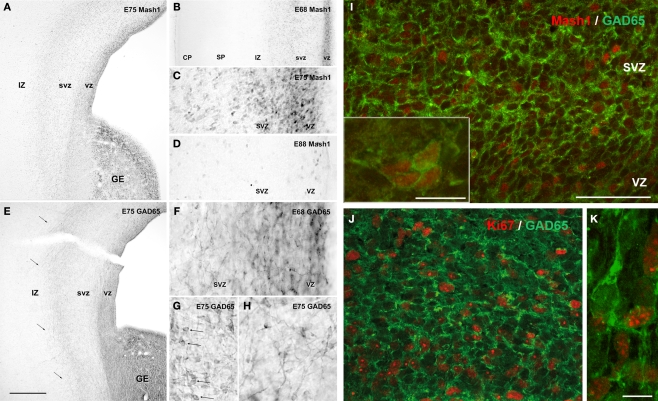
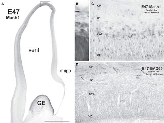
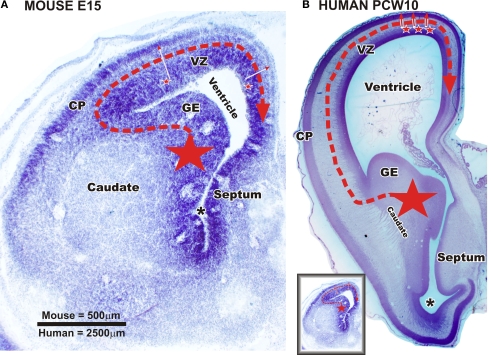
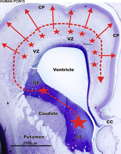
Gamma-aminobutyric-acidergic (GABAergic) cells form a very heterogeneous population of neurons that play a crucial role in the coordination and integration of cortical functions. Their number and diversity increase through mammalian brain evolution. Does evolution use the same or different developmental rules to provide the increased population of cortical GABAergic neurons? In rodents, these neurons are not generated in the pallial proliferative zones as glutamatergic principal neurons. They are produced almost exclusively by the subpallial proliferative zones, the ganglionic eminence (GE) and migrate tangentially to reach their target cortical layers. The GE is organized in molecularly different subdomains that produce different subpopulations of cortical GABAergic neurons. In humans and non-human primates, in addition to the GE, cortical GABAergic neurons are also abundantly generated by the proliferative zones of the dorsal telencephalon. Neurogenesis in ventral and dorsal telencephalon occurs with distinct temporal profiles. These dorsal and ventral lineages give rise to different populations of GABAergic neurons. Early-generated GABAergic neurons originate from the GE and mostly migrate to the marginal zone and the subplate. Later-generated GABAergic neurons, originating from both proliferative sites, populate the cortical plate. Interestingly, the pool of GABAergic progenitors in dorsal telencephalon produces mainly calretinin neurons, a population known to be significantly increased and to display specific features in primates. We conclude that the development of cortical GABAergic neurons have exclusive features in primates that need to be considered in order to understand pathological mechanisms leading to some neurological and psychiatric diseases.

Keywords: ganglionic eminence; glutamic acid decarboxylase; interneurons; neurogenesis; tangential migration; ventricular zone.


Figures

Figure 1
Figure 1
Origin and migratory routes differ between the two major classes of cortical neurons. (A) Shematic drawing of migratory routes for cortical neurons in the dorsal telencephalon (pallium). Principal (pyramidal) neurons (excitatory glutamatergic neurons) originate locally in the proliferative ventricular (VZ) and subventricular (SVZ) zones of the dorsal telencephalon. Early-generated neurons display a bipolar morphology and migrate orthogonal to the surface of the brain along radially oriented glial fibres (radial migration). According to data obtained in rodents, hippocampal and cortical GABAergic neurons originate in the ventral proliferative zones, i.e. ganglionic eminence (GE) and migrate tangentially to their target cortical regions (red arrowed lines). The largest stream follows the lower intermediate zone (IZ)/upper subventricular zone (SVZ). In the marginal zone (MZ), the stream of migrating neurons forms a subpial granular layer. Numerous tangentially oriented neurons are present below the cortical plate (CP), in the subplate. Migratory routes are shown on schematized frontal section of human fetus at postconceptual week 15 (Th – thalamus; C – caudate nucleus; P – putamen; CC – corpus callosum). (B) Microphotography of a Golgi impregnated section through the whole thickness of dorsal telencephalon in a human fetus at postconceptual week 10. Bipolar migrating, prospective principal neurons are mostly observed in the VZ and SVZ. Numerous radial glia cells extend their processes from ventricle to pia. Numerous prospective pyramidal neurons at the beginning of differentiation are present in the cortical plate. Red arrows indicate major locations and direction of tangential migration. Modified by permission from Kostovic et al. (2007).
Figure 2
Figure 2
Neurogenesis of GABAergic neurons in the ventral telencephalon of a monkey fetus at embryonic day (E) 55. (A,B) Adjacent sections labeled for Mash1 (A) and GAD65 (B) from a E55 fetal brain at a rostral level in which the lateral ventricle is dilated (paleocortical ventricle indicated by a star). (A) Progenitors labeled for Mash1 were present mainly in the ganglionic eminence (GE) [box magnified in panel (C)] and septal (Sept) proliferative zone [box magnified in panel (D)]. (B) GAD65-containing cells were observed in the same regions of the ventral telencephalon as Mash1, the GE and the Sept [boxes enlarged in panels (E) and (F)]. (C–F) Figures correspond to magnified regions indicated by boxes in (A,B). (C) In the GE many cells were labeled for Mash1 in the ventricular (VZ) and the subventricular zone (SVZ). The intensity of labeling was much higher in the VZ than in the SVZ. (D) In the prospective septal region (Sept), numerous cells highly labeled for Mash1 were observed in the VZ whereas the cells in the SVZ displayed a moderate level of labeling. (E) Many cells labeled for GAD65 were observed in the GE. These cells were mainly located in the SVZ, and only a few cells were present in the VZ. (F) Many cells labeled for GAD65 were present in the SVZ of the Sept. Box: magnified region. (G) Confocal micrographs of an adjacent coronal section double-labeled for Mash1 (red, nuclear) and GAD65 (green, cytoplasmic) showing that in the SVZ of GE numerous Mash1 containing progenitors were labeled for GAD65. Scale bars: 1 mm (A,B), 50 μm (C–G), 10 μm [box in (F)]. Abbreviation: CC – corpus callosum. Modified by permission from Petanjek et al. (2009).
Figure 3
Figure 3
Tangential migration of GABAergic neurons in coronal sections of a monkey fetus at embryonic day (E) 55. (A) Mash1 containing progenitors were present in the ganglionic eminence (GE) but not in the neighboring structures including the caudate nucleus (caud), capsula interna (CI), and adjacent zones of the dorsal telencephalon: ventricular zone (VZ), subventricular zone (SVZ) and intermediate zone (IZ). Arrows indicated the border between GE and adjacent structures. (B) GAD65 containing progenitors were present almost exclusively in the GE. In addition a stream of post-mitotic-like, GAD65-containing neurons extending from the GE to dorsal telencephalon was observed in the lower IZ/upper SVZ. Arrows indicate the deeper border of this stream. Boxes: magnified regions in panels (C) and (D). (C) GAD65-containing cells with a characteristic morphology of post-mitotic migrating neurons, including a leading process (arrows) leaving the GE. (D) In the IZ and upper SVZ of dorsal telencephalon, the majority of GAD65 migrating neurons were tangentially oriented (arrows). (E) Tangentially oriented GAD65 migrating neurons are also observed in the subplate (SP) (arrows), cortical plate (CP) and marginal zone (MZ) of the dorsal telencephalon. (F,G) Confocal micrographs of an adjacent coronal section double-labeled for Ki67 (F) or Mash1 (G) (red, nuclear) and GAD65 (green, cytoplasmic). At E55, in the SVZ of the GE many GAD65-labeled cells were labeled for Ki67 (F) and therefore corresponded to proliferating cells. In contrast none of the migrating GAD65-containing cells in the IZ displayed detectable level of Ki67 or Mash1 (G) and therefore likely corresponded to early post-mitotic neurons. Scale bars: 350 μm (A,B), 50 μm (F,G), 20 μm, (C–E). Modified by permission from Petanjek et al. (2009).
Figure 4
Figure 4
Neurogenesis of GABAergic neurons in the ventral and dorsal telencephalon of monkey fetuses at embryonic day (E) 68, E75 and E88. (A–D) Mash1 expression. (A) At E75, Mash1 containing progenitors were present in the ganglionic eminence (GE) as well as in ventricular (VZ) and subventricular (SVZ) zone of the entire dorsal telencephalon. In both areas, the intensity of labeling for Mash1 decreased from the VZ to the SVZ. (B) At E68, the same pattern of labeling for Mash1 is already present in the VZ and SVZ of the dorsal telencephalon. No labeling is observed in the upper part of intermediate zone (IZ), the subplate (SP) and the cortical plate (CP). (C) Higher magnification of panel (A). The numerous Mash1 highly labeled cells located in the VZ and SVZ of the dorsal telencephalon. (D) At E88, end of neurogenesis, the intensity of labeling for Mash1 was strongly decreased as compared to E68 (B) and E75 (A,C). (E–H) GAD65 expression. (E) At E75, GAD65-containing progenitors were present in the GE, but also in the proliferative VZ and SVZ of the dorsal telencephalon in contrast to earlier stages. The stream of migrating GAD65-containing neurons observed at E55 [compare with panel (B) in Figure 3] in the lower part of IZ and upper part of SVZ was still present (arrows). (F) At E68, similar labeling was already observed. In contrast to previous stages many migrating-like post-mitotic neurons with one leading process were now observed in the VZ and SVZ of the dorsal telencephalon. (G) At E75, in addition to migrating-like post-mitotic neurons, many round-shaped cells without any process were labeled for GAD65 (arrows). These cells displayed a similar morphology to that observed in the GE and septal region. (H) At E75, many migrating-like GAD65-neurons in the SVZ of the dorsal telencephalon displayed multidirectional orientations. (I–J) Confocal micrographs of an adjacent coronal section double-labeled for Mash1 (I) or Ki67 (J,K) (red, nuclear) and GAD65 (green, cytoplasmic). (I) At E75, in the dorsal telencephalon, most Mash1-containing cells of SVZ also contained GAD65, whereas many Mash1-labeled cells in the VZ did not. Insert: aggregates of cells double-labeled for Mash1 and GAD65 were frequently observed in the SVZ of the dorsal telencephalon, suggesting cells undergoing division. (J) At E75 in the SVZ of the dorsal telencephalon [compare with panel (G)] most GAD65-containing cells were labeled for Ki67 and therefore corresponded to progenitors of GABAergic neurons. (K) Migrating-like GAD65-containing cells in the IZ of the dorsal telencephalon were not labeled for Ki67. Scale bars: 550 μm (A,E), 250 μm (B), 60 μm (C,D) and 30 μm (F–H); 50 μm (I,J), 10 μm [insert (I,K)]. Modified by permission from Petanjek et al. (2009).
Figure 5
Figure 5
Mash1 and GAD65 labeling patterns in coronal sections of monkey fetal telencephalon at embryonic day (E) 47 suggest that neurogenesis of GABAergic neurons is induced locally. (A) At E47 Mash1 was highly expressed in the proliferative zone of the ventral telencephalon, the ganglionic eminence (GE). Mash1 immunoreactivity was also observed in the proliferative zones of the dorsal telencephalon. It followed a clear dorso-ventral decreasing gradient in the labeling intensity within the entire cortical wall. Moderate level of labeling was observed in the most dorsal part of the telencephalon [indicated by square and magnified in panel (C)]. The intensity of staining decreased in the lateral and medial wall. No Mash1 immunolabeling was observed close to the GE, nor in the most ventral part of the medial telencephalic wall, that corresponds at this level to dorsal hippocampus (dhipp). (B) Cresyl violet labeled section. Different cellular zones of the most dorsal part of the dorsal telencephalon are delineated. (C) Some cells moderately labeled for Mash1 were observed in the proliferative zones of the most dorsal part of the dorsal telencephalon. Most of these Mash1 labeled cells were present in the upper part of subventricular zone (SVZ), on the border with the intermediate zone (IZ). Some cells were also observed in the deepest part of ventricular zone (VZ). (D) In contrast to Mash1 no GAD65-containing cells were observed in the VZ and SVZ of the most dorsal part of the dorsal telencephalon at E47. However in this region, some GAD65-containing cells with a characteristic morphology of early post-mitotic migrating neurons, including a leading process, and tangentially oriented, were observed in the IZ and the cortical plate (CP) (arrows). Vent – lateral ventricle. Scale bars: 1 mm (A), 50 μm (B–D). Modified by permission from Petanjek et al. (2009).
Figure 6
Figure 6
Sources of cortical GABAergic neuron in a mouse fetus and in primate fetuses at early developmental stage (embryonic day (E) 47 to E55 in macaque/postconceptional week (PCW) 10 to PCW13 in human). Nissl staining of frontal brain sections through telencephalic vesicle at the level of septum and paleocortical ventricle (asterix) in a mouse fetus at embryonic day (E) 15 [panel (A) and in a human fetus at postconceptional week 10 panel (B)]. (A) In the rodent, the vast majority of cortical and hippocampal GABAergic neurons are produced by ganglionic eminence (GE) (large star). The neurons migrate tangentially in the cortex (red dashed arrow). A very small percentage of the GABAergic neurons is supposed to be also produced in dorsal proliferative zones (small stars – full red arrows). (B) In the monkey and human brain, the GE is also an important source of cortical GABAergic neurons (large star). Post-mitotic neurons migrate tangentially into the cortex (red dashed arrow). Data from macaque monkey (Petanjek et al., 2009) suggest that already at this ages (see below) a production of GABAergic neurons started in the proliferative zones of most dorsal part of the pallium (small stars – full red arrows). VZ – ventricular and subventricular zone; CP – cortical plate. Scale bar: 500 μm (A), 2500 μm (B). Small figure [left down in panel (B)] is a mouse section at E15 (A) reduced in size to be comparable with human fetus at PCW10 (B).
Figure 7
Figure 7
Sources of cortical GABAergic neuron in the the primate during middle fetal stage. Nissl staining of frontal brain sections through rostral part of the telencephalic vesicle in a human fetus at postconceptional week (PCW) 15 (at a level of the anterior horn). In human (PCW15 – PCW20) as in macaque monkey [embryonic day (E) 64 to E75] later during gestation, GABAergic neurons are produced massively by the proliferative zones of the dorsal telencephalon (smaller stars – full red arrows). According to Letinic et al. (2002) these dorsal proliferatives zones generate 65% of the cortical GABAergic neurons. This massive dorsal production of GABAergic neurons is primate-specific. VZ – ventricular and subventricular zone; CP – cortical plate; CC – corpus callosum. Scale bar: 2500 μm.

Similar articles

Cited by

References

    1. Anderson S. A., Eisenstat D. D., Shi L., Rubenstein J. L. (1997). Interneuron migration from basal forebrain to neocortex: dependence on Dlx genes. Science 278, 474–47610.1126/science.278.5337.474 - DOI - PubMed
    1. Anderson S. A., Kaznowski C. E., Horn C., Rubenstein J. L., McConnell S. K. (2002). Distinct origins of neocortical projection neurons and interneurons in vivo. Cereb. Cortex 12, 702–70910.1093/cercor/12.7.702 - DOI - PubMed
    1. Andrioli A., Alonso-Nanclares L., Arellano J. I., DeFelipe J. (2007). Quantitative analysis of parvalbumin-immunoreactive cells in the human epileptic hippocampus. Neuroscience 149, 131–14310.1016/j.neuroscience.2007.07.029 - DOI - PubMed
    1. Ang E. S., Jr, Haydar T. F., Gluncic V., Rakic P. (2003). Four-dimensional migratory coordinates of GABAergic interneurons in the developing mouse cortex. J. Neurosci. 23, 5805–5815 - PMC - PubMed
    1. Ascoli G. A., Alonso-Nanclares L., Anderson S. A., Barrionuevo G., Benavides-Piccione R., Burkhalter A., Buzsaki G., Cauli B., Defelipe J., Fairen A., Feldmeyer D., Fishell G., Fregnac Y., Freund T. F., Gardner D., Gardner E. P., Goldberg J. H., Helmstaedter M., Hestrin S., Karube F., Kisvarday Z. F., Lambolez B., Lewis D. A., Marin O., Markram H., Munoz A., Packer A., Petersen C. C., Rockland K. S., Rossier J., Rudy B., Somogyi P., Staiger J. F., Tamas G., Thomson A. M., Toledo-Rodriguez M., Wang Y., West D. C., Yuste R. (2008). Petilla terminology: nomenclature of features of GABAergic interneurons of the cerebral cortex. Nat. Rev. Neurosci. 9, 557–56810.1038/nrn2402 - DOI - PMC - PubMed

LinkOut - more resources