

Dependence of zonal chondrocyte water transport properties on osmotic environment
- PMID: 20011231
- PMCID: PMC2792913
- DOI: 10.1007/s12195-008-0026-6
Dependence of zonal chondrocyte water transport properties on osmotic environment
Abstract
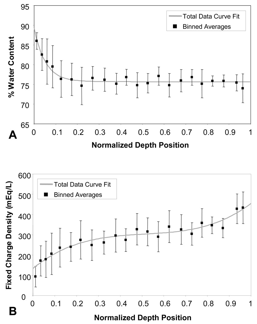
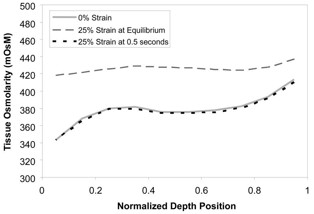
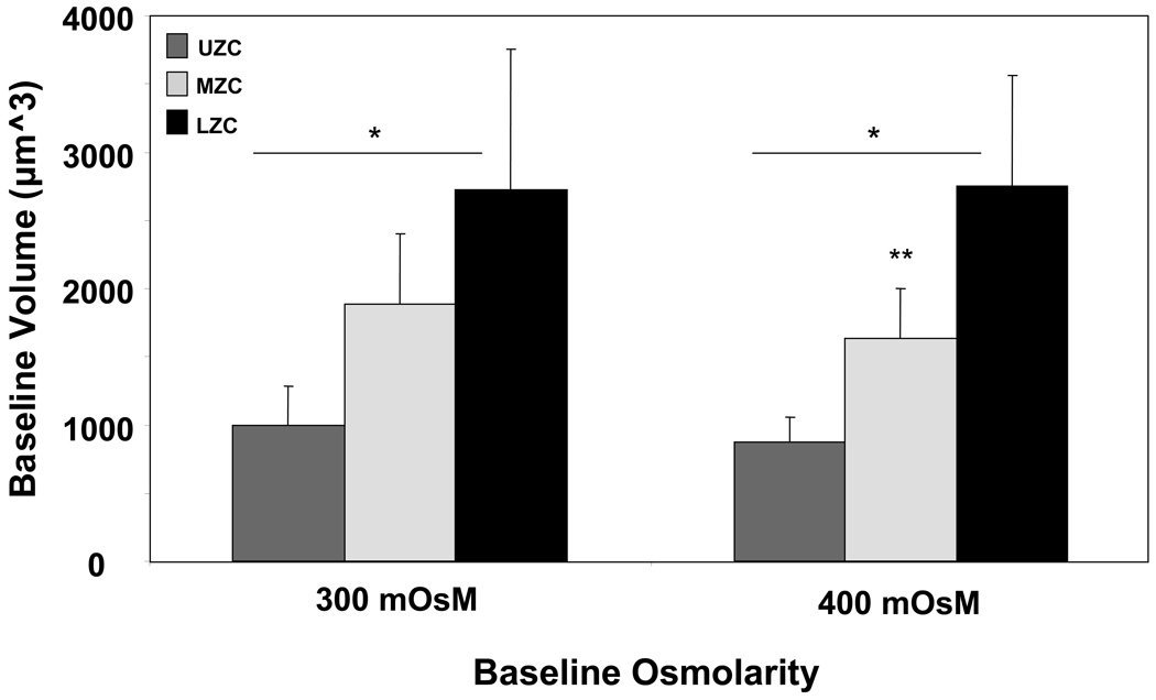
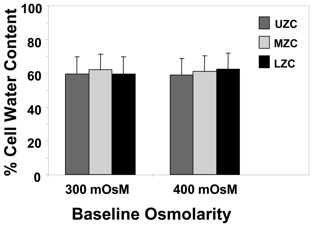
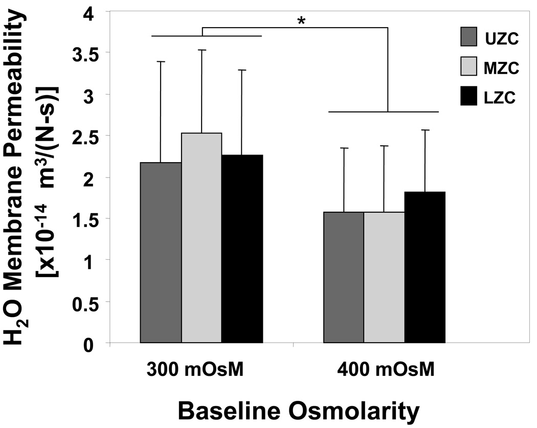
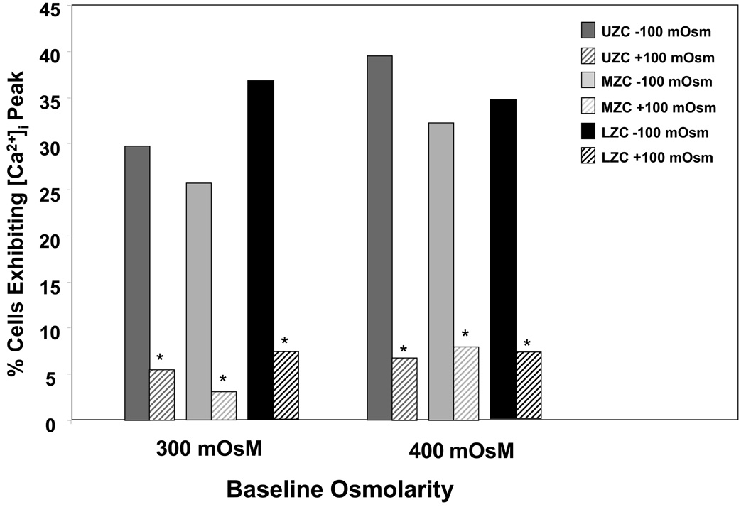
OBJECTIVE: The increasing concentration of proteoglycans from the surface to the deep zone of articular cartilage produces a depth-dependent gradient in fixed charge density, and therefore extracellular osmolarity, which may vary with loading conditions, growth and development, or disease. In this study we examine the relationship between in situ variations in osmolarity on chondrocyte water transport properties. Chondrocytes from the depth-dependent zones of cartilage, effectively preconditioned in varying osmolarities, were used to probe this relationship. DESIGN: First, depth variation in osmolarity of juvenile bovine cartilage under resting and loaded conditions was characterized using a combined experimental/theoretical approach. Zonal chondrocytes were isolated into two representative "baseline" osmolarities chosen from this analysis to reflect in situ conditions. Osmotic challenge was then used as a tool for determination of water transport properties at each of these baselines. Cell calcium signaling was monitored simultaneously as a preliminary examination of osmotic baseline effects on cell signaling pathways. RESULTS: Osmotic baseline exhibits a significant effect on the cell membrane hydraulic permeability of certain zonal subpopulations but not on cell water content or incidence of calcium signaling. CONCLUSIONS: Chondrocyte properties can be sensitive to changes in baseline osmolarity, such as those occurring during OA progression (decrease) and de novo tissue synthesis (increase). Care should be taken in comparing chondrocyte properties across zones when cells are tested in vitro in non-physiologic culture media.
Figures
Similar articles
-
Osmolarity influences chondrocyte death in wounded articular cartilage.J Bone Joint Surg Am. 2008 Jul;90(7):1531-42. doi: 10.2106/JBJS.G.00857. J Bone Joint Surg Am. 2008. PMID: 18594103
-
Passive osmotic properties of in situ human articular chondrocytes within non-degenerate and degenerate cartilage.J Cell Physiol. 2005 Jul;204(1):309-19. doi: 10.1002/jcp.20294. J Cell Physiol. 2005. PMID: 15668989
-
The osmotic sensitivity of isolated and in situ bovine articular chondrocytes.J Orthop Res. 2001 Sep;19(5):768-78. doi: 10.1016/S0736-0266(01)00013-4. J Orthop Res. 2001. PMID: 11562120
-
Cartilage regeneration using zonal chondrocyte subpopulations: a promising approach or an overcomplicated strategy?J Tissue Eng Regen Med. 2015 Jun;9(6):669-78. doi: 10.1002/term.1638. Epub 2012 Nov 8. J Tissue Eng Regen Med. 2015. PMID: 23135870 Review.
-
Perspective in Achieving Stratified Articular Cartilage Repair Using Zonal Chondrocytes.Tissue Eng Part B Rev. 2023 Jun;29(3):310-330. doi: 10.1089/ten.TEB.2022.0142. Epub 2023 Jan 24. Tissue Eng Part B Rev. 2023. PMID: 36416231 Review.
Cited by
-
Label-free protein profiling of adipose-derived human stem cells under hyperosmotic treatment.J Proteome Res. 2011 Jul 1;10(7):3050-9. doi: 10.1021/pr200030v. Epub 2011 Jun 14. J Proteome Res. 2011. PMID: 21604804 Free PMC article.
-
Importance of Osmolarity and Oxygen Tension for Cartilage Tissue Engineering.Biores Open Access. 2020 Mar 31;9(1):106-115. doi: 10.1089/biores.2020.0009. eCollection 2020. Biores Open Access. 2020. PMID: 32257626 Free PMC article.
-
Applied osmotic loading for promoting development of engineered cartilage.J Biomech. 2013 Oct 18;46(15):2674-81. doi: 10.1016/j.jbiomech.2013.07.043. Epub 2013 Aug 30. J Biomech. 2013. PMID: 24035014 Free PMC article.
-
Physicochemical and biomechanical stimuli in cell-based articular cartilage repair.Curr Rheumatol Rep. 2015 Mar;17(3):22. doi: 10.1007/s11926-014-0493-9. Curr Rheumatol Rep. 2015. PMID: 25828845 Free PMC article. Review.
-
Trimethylamine N-oxide as a media supplement for cartilage tissue engineering.J Orthop Res. 2012 Dec;30(12):1898-905. doi: 10.1002/jor.22171. Epub 2012 Jun 15. J Orthop Res. 2012. PMID: 22707357 Free PMC article.
References
-
- Aydelotte MB, Greenhill RR, Kuettner KE. Differences between sub-populations of cultured bovine articular chondrocytes. II. Proteoglycan metabolism. Connect Tissue Res. 1988;18:223–234. - PubMed
Grants and funding
LinkOut - more resources
Full Text Sources
