. 2009 Dec 11;4(12):e8262.
doi: 10.1371/journal.pone.0008262.

The 3'-phosphoadenosine 5'-phosphosulfate transporters, PAPST1 and 2, contribute to the maintenance and differentiation of mouse embryonic stem cells

Affiliations

The 3'-phosphoadenosine 5'-phosphosulfate transporters, PAPST1 and 2, contribute to the maintenance and differentiation of mouse embryonic stem cells

Norihiko Sasaki et al. PLoS One. .

Abstract

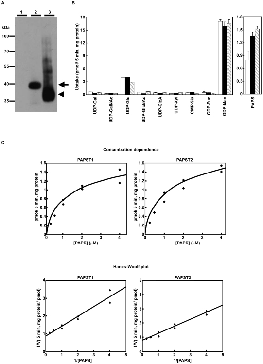
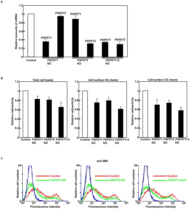
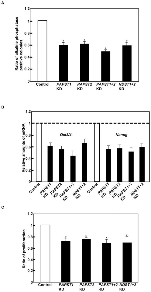
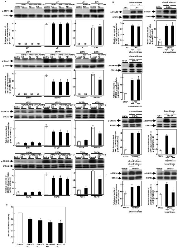
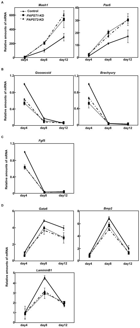
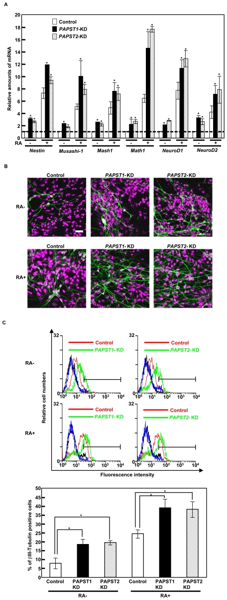
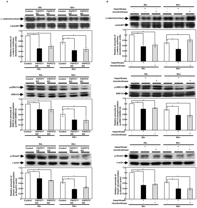
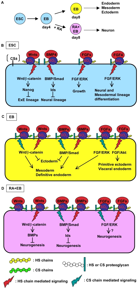
Recently, we have identified two 3'-phosphoadenosine 5'-phosphosulfate (PAPS) transporters (PAPST1 and PAPST2), which contribute to PAPS transport into the Golgi, in both human and Drosophila. Mutation and RNA interference (RNAi) of the Drosophila PAPST have shown the importance of PAPST-dependent sulfation of carbohydrates and proteins during development. However, the functional roles of PAPST in mammals are largely unknown. Here, we investigated whether PAPST-dependent sulfation is involved in regulating signaling pathways required for the maintenance of mouse embryonic stem cells (mESCs), differentiation into the three germ layers, and neurogenesis. By using a yeast expression system, mouse PAPST1 and PAPST2 proteins were shown to have PAPS transport activity with an apparent K(m) value of 1.54 microM or 1.49 microM, respectively. RNAi-mediated knockdown of each PAPST induced the reduction of chondroitin sulfate (CS) chain sulfation as well as heparan sulfate (HS) chain sulfation, and inhibited mESC self-renewal due to defects in several signaling pathways. However, we suggest that these effects were due to reduced HS, not CS, chain sulfation, because knockdown of mouse N-deacetylase/N-sulfotransferase, which catalyzes the first step of HS sulfation, in mESCs gave similar results to those observed in PAPST-knockdown mESCs, but depletion of CS chains did not. On the other hand, during embryoid body formation, PAPST-knockdown mESCs exhibited abnormal differentiation, in particular neurogenesis was promoted, presumably due to the observed defects in BMP, FGF and Wnt signaling. The latter were reduced as a result of the reduction in both HS and CS chain sulfation. We propose that PAPST-dependent sulfation of HS or CS chains, which is regulated developmentally, regulates the extrinsic signaling required for the maintenance and normal differentiation of mESCs.


Conflict of interest statement

Competing Interests: The authors have declared that no competing interests exist.

Figures

Figure 1
Figure 1. Both mouse PAPST1 and PAPST2 encode PAPS transporter proteins.
(A) Expression state of PAPST1 and PAPST2 proteins in the Golgi-enriched fraction. Western blot analysis of the P100 fractions prepared from yeast cells expressing either the mock vector (lane 1), HA-tagged PAPST1 (lane 2) or HA-tagged PAPST2 (lane 3). An aliquot of 5 µg of protein from the control cells and cells expressing HA-tagged PAPST1 or 0.5 µg of protein from the cells expressing HA-tagged PAPST2 was loaded. The arrow and arrowhead indicate HA-tagged PAPST1 and HA-tagged PAPST2, respectively. (B) Substrate specificity of PAPST1 and PAPST2. Each P100 fraction was incubated in 50 µl of reaction buffer containing 1 µM labelled substrate at 32°C for 5 min, and the radioactivity incorporated was measured. The indicated values are the mean±SD obtained from two independent experiments (open bars, Mock; solid bars, PAPST1; hatched bars, PAPST2). (C) Substrate concentration dependence. Each P100 fraction was incubated in 50 µl of reaction buffer containing different concentrations of [35S]PAPS at 32°C for 5 min, and the radioactivity incorporated was measured. Specific incorporation was calculated by subtracting the value for the mock transfection from each of the values obtained. Lower panel, the Hanes-Woolf plot used to determine the Km value is shown.
Figure 2
Figure 2. Knockdown of PAPST1 or PAPST2 mRNA induced reduction of sulfation in mESCs.
(A) Real time PCR analysis of cells 2 days after transfection. Relative amounts of PAPST mRNA were calculated after normalization to β-actin mRNA in the same cDNA. The results are shown after normalization against the values obtained with control cells (value = 1). The values shown are the means±SD of three independent experiments. (B) Metabolic labeling analysis. The results of total sulfate incorporation into cellular proteins and sulfate incorporation into cell surface HS and CS chains are shown after normalization against the values obtained with control cells (value = 1). The values shown are the means±SD of three independent experiments and significant values are indicated; *P<0.01, in comparison to the control. (C) FACS analysis of cells 3 days after transfection using an anti-SM3 antibody (black and blue lines represent the IgM isotype control for control and PAPST-KD cells, respectively). Three independent experiments were performed and representative results are shown.
Figure 3
Figure 3. Both PAPST1- and PAPST2-KD cells showed decreased potential for self-renewal and proliferation.
(A) Self-renewal assay. The ratio of alkaline phosphatase positive colonies is shown after normalization against the ratio obtained with control cells (value = 1). Approximately 70% of the colonies derived from the control cells remained in an undifferentiated state in feeder-free culture. The values shown are the means±SD from three independent experiments and significant values are indicated; *P<0.01, in comparison to the control. (B) Real time PCR analysis of undifferentiated state markers in the cells 4 days after transfection. The results are shown after normalization against the values obtained for control cells (value = 1). The values shown are the means±SD from two independent experiments. (C) Proliferation assay. The ratio of proliferation 48 h after culture is shown after normalization against the values obtained with control cells (value = 1). The values shown are the means±SD from three independent experiments and significant values are indicated; *P<0.01, in comparison to the control.
Figure 4
Figure 4. Signaling by specific factors was decreased in PAPST-KD cells, but not in CS chain-depleted cells.
(A) and (B) Western blot analysis of cells stimulated with the extrinsic factors. Cell lysate was prepared as described in Materials and Methods. Two independent experiments were performed and representative results are shown. The histograms show mean densitometric readings±SD of the phosphorylated protein/loading controls. Values were obtained from duplicate measurements of two independent experiments and significant values are indicated; *P<0.05, in comparison to the stimulated control; ND, not detected. (C) Luciferase reporter assay. Relative luciferase activities (TOPFLASH/FOPFLASH) are shown as means±SD from three independent experiments after normalization against the values obtained with control cells (value = 1), and significant values are indicated; *P<0.05, in comparison to the control.
Figure 5
Figure 5. Abnormal differentiation was observed in PAPST-KD cells during EB formation.
(A)–(D) Real time PCR analysis of germ layer markers 4, 8, and 12 days after EB formation (A, neurectoderm marker; B, mesoderm marker; C, primitive ectoderm marker; D, extraembryonic endoderm (ExE) marker). The results are shown after normalization against the values obtained with control EBs on day 4 (value = 1). The values shown are the means±SD of two independent experiments.
Figure 6
Figure 6. Neurogenesis was promoted in PAPST-KD cells.
(A) Real time PCR analysis of neural differentiation markers 8 days after EB formation. The results are shown after normalization against the values obtained with control cells not treated with RA (value = 1). The values shown are the means±SD of duplicate measurements from two independent experiments and significant values are indicated; *P<0.05, in comparison to the control. (B) Immunocytochemical staining 6 days after replating of EBs. Representative confocal images from two independent experiments are shown. (βIII-Tubulin, green; PI, purple). Scale bar, 20 µm. (C) FACS analysis 6 days after replating of EBs using an anti-βIII-Tubulin antibody (black and blue lines represent the IgM isotype control for control and PAPST-KD cells, respectively). Three independent experiments were performed and representative results are shown. The histograms show the ratio of the mean fluorescent intensity within area of the insetted bar representing βIII-Tubulin positive cells to the mean fluorescent intensity over the total area±SD of three independent experiments and significant values are indicated; *P<0.01, in comparison to the control.
Figure 7
Figure 7. Signaling via a number of pathways was decreased in PAPST-KD cells during EB formation.
(A) Western blot analysis of several signaling molecules in EBs on day 8. Two independent experiments were performed and representative results are shown. The histograms show mean densitometric readings±SD of β-catenin or the phosphorylated proteins/loading controls after normalization against the values obtained with control cells not treated with RA (value = 1). Values were obtained from duplicate measurements of two independent experiments and significant values are indicated; *P<0.01, in comparison to the control. (B) Western blot analysis of several signaling molecules in EBs on day 8 after heparitinase or chondroitinase treatment. Two independent experiments were performed and representative results are shown. The histograms show mean densitometric readings±SD of β-catenin or the phosphorylated proteins/loading controls after normalization against the values obtained with cells not treated with RA and enzyme (value = 1). Values were obtained from duplicate measurements of two independent experiments and significant values are indicated; *P<0.01, in comparison to cells not treated with enzyme.
Figure 8
Figure 8. PAPST-dependent sulfation contributes to mESCs in both undifferentiated and differentiated state.
(A) In vitro differentiation flowchart of mESCs. EBs that are not treated with RA produce cells from all three germ layers (endoderm, mesoderm and ectoderm) whereas RA-treated EBs produce neurons after further adherent culture. (B) PAPST-dependent sulfation of HS chains regulates the extrinsic signaling (by BMP, FGF and Wnt) that is required for the growth and pluripotency of mESCs. In undifferentiated mESCs, the transduction of extrinsic signals is dependent on the sulfation of HS chains, but not CS chains, and this maintains growth and pluripotency. As shown in this study, autocrine/paracrine FGF signaling contributes to the growth of mESCs. In particular, FGF4 signaling contributes to the differentiation state of the mESCs . (C) PAPST-dependent sulfation of both HS and CS chains regulates the extrinsic signaling (by BMP, FGF and Wnt) that is required for normal differentiation of EBs. During EB differentiation into the three germ layers, the transduction of the extrinsic signals is dependent on the sulfation of both HS and CS chains. Wnt and BMP signaling inhibit ectodermal differentiation and contribute to mesodermal and definitive endodermal differentiation –, , , . FGF/ERK and FGF/Akt signaling contribute to mesodermal and definitive endodermal differentiation and primitive ectodermal and visceral endodermal differentiation, respectively , , . (D) PAPST-dependent sulfation of HS and CS chains regulates the extrinsic signaling (by BMP, Wnt and FGF) that is required for neuronal differentiation of RA-treated EBs. During RA-treated EB differentiation, the transduction of extrinsic signals is dependent on the sulfation of both HS and CS chains and results in neuronal differentiation. Wnt and BMP signaling inhibit neurogenesis , and FGF (e.g., bFGF) signaling may contribute to neurogenesis. CS chains regulate Wnt signaling negatively, presumably by sequestering Wnt proteins and preventing them interacting with Wnt receptors.

Similar articles

Cited by

References

    1. Evans MJ, Kaufman MH. Establishment in culture of pluripotential cells from mouse embryos. Nature. 1981;292:154–156. - PubMed
    1. Martin GR. Isolation of a pluripotent cell line from early mouse embryos cultured in medium conditioned by teratocarcinoma stem cells. Proc Natl Acad Sci U S A. 1981;78:7634–7638. - PMC - PubMed
    1. Keller G. Embryonic stem cell differentiation: emergence of a new era in biology and medicine. Genes Dev. 2005;19:1129–1155. - PubMed
    1. Chambers I, Smith A. Self-renewal of teratocarcinoma and embryonic stem cells. Oncogene. 2004;23:7150–7160. - PubMed
    1. Ivanova N, Dobrin R, Lu R, Kotenko I, Levorse J, et al. Dissecting self-renewal in stem cells with RNA interference. Nature. 2006;442:533–538. - PubMed

Publication types

MeSH terms