. 2009 Dec;5(12):e1000691.
doi: 10.1371/journal.ppat.1000691. Epub 2009 Dec 11.

Inhibition of adaptive immune responses leads to a fatal clinical outcome in SIV-infected pigtailed macaques but not vervet African green monkeys

Affiliations

Inhibition of adaptive immune responses leads to a fatal clinical outcome in SIV-infected pigtailed macaques but not vervet African green monkeys

Jörn E Schmitz et al. PLoS Pathog. 2009 Dec.

Abstract

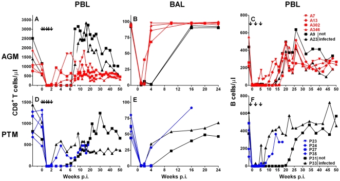
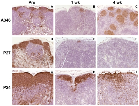
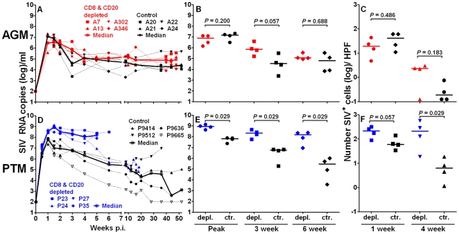
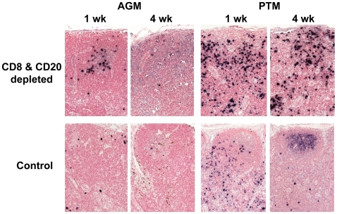
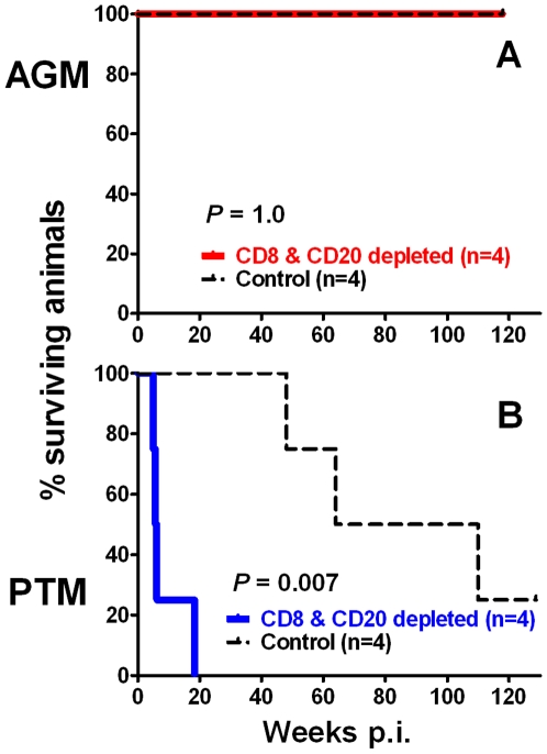
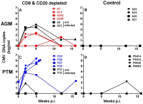
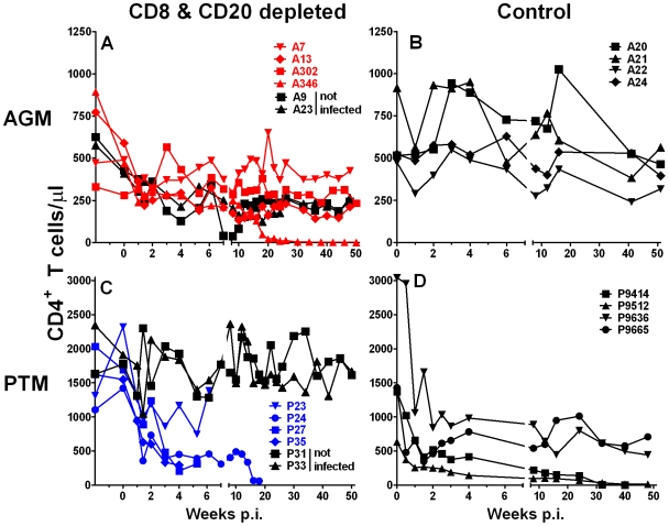
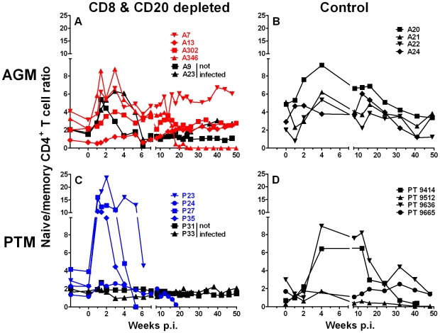
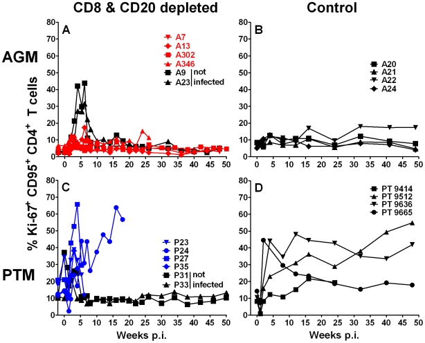
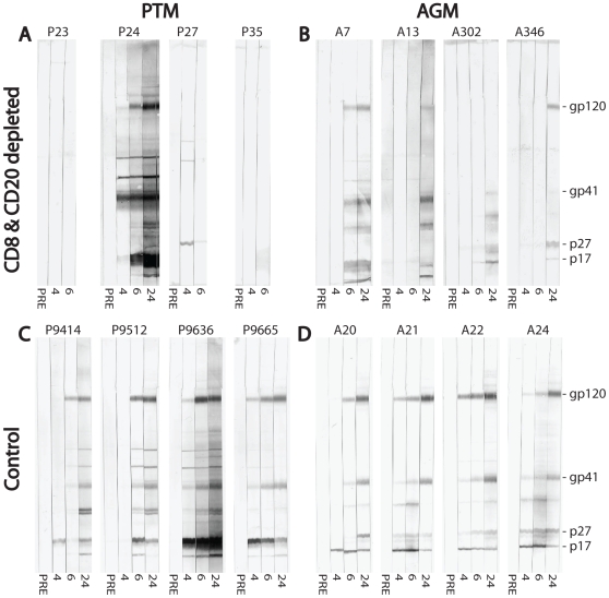
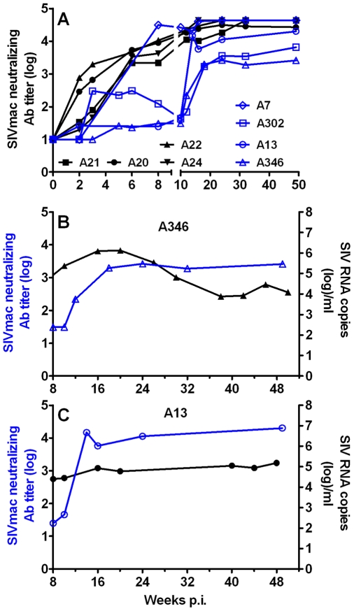
African green monkeys (AGM) and other natural hosts for simian immunodeficiency virus (SIV) do not develop an AIDS-like disease following SIV infection. To evaluate differences in the role of SIV-specific adaptive immune responses between natural and nonnatural hosts, we used SIV(agmVer90) to infect vervet AGM and pigtailed macaques (PTM). This infection results in robust viral replication in both vervet AGM and pigtailed macaques (PTM) but only induces AIDS in the latter species. We delayed the development of adaptive immune responses through combined administration of anti-CD8 and anti-CD20 lymphocyte-depleting antibodies during primary infection of PTM (n = 4) and AGM (n = 4), and compared these animals to historical controls infected with the same virus. Lymphocyte depletion resulted in a 1-log increase in primary viremia and a 4-log increase in post-acute viremia in PTM. Three of the four PTM had to be euthanized within 6 weeks of inoculation due to massive CMV reactivation and disease. In contrast, all four lymphocyte-depleted AGM remained healthy. The lymphocyte-depleted AGM showed only a trend toward a prolongation in peak viremia but the groups were indistinguishable during chronic infection. These data show that adaptive immune responses are critical for controlling disease progression in pathogenic SIV infection in PTM. However, the maintenance of a disease-free course of SIV infection in AGM likely depends on a number of mechanisms including non-adaptive immune mechanisms.


Conflict of interest statement

The authors have declared that no competing interests exist.

Figures

Figure 1
Figure 1. CD8+ T cell and CD20+ B cell depletion in AGM and PTM.
Anti-CD8 antibody administration-induced depletion of CD8+ lymphocytes in peripheral blood lymphocytes (PBL) and bronchoalveolar lavage (BAL) in AGM (A, B) and PTM (D, E). Anti-CD20 antibody administration induced depletion of B cells in PBL of vervet AGM (C) and of PTM (F). Animals that received anti-CD8 and anti-CD20 antibodies but were not inoculated with SIV are shown with black symbols. Animals that received anti-CD8 and anti-CD20 antibodies and were inoculated with SIVagmVer90 are shown in color symbols (red for AGM, blue for PTM). The black arrows in panels A and D indicate the injection of the anti-CD8α mAb cM-T807. The black arrows in panels C and F indicate the injection of the anti-CD20 mAb Rituximab.
Figure 2
Figure 2. Only partial depletion of B cells in lymphatic tissues of CD8+ and CD20+ lymphocyte-depleted AGM but not of PTM.
Immunohistochemical (IHC) detection of CD20+ B cells in sequential lymph node biopsies from a representative vervet AGM (A346) collected pre-inoculation (A), one week p.i. (B), and four weeks p.i. (C). Representative IHC staining for CD20 is shown on two PTM with different clinical courses. PTM P27 was representative of the pattern seen in the three PTM that showed efficient depletion of CD20+ B cells at one week p.i. that was irreversible at four weeks p.i. (D, E, and F). PTM P24 only had a partial depletion of CD20+ B cells at one week p.i. and near normal CD20+ B cell levels at four weeks p.i. (G, H, and I).
Figure 3
Figure 3. CD8+ and CD20+ lymphocyte depletion has a greater effect on SIV peak viremia and lymphatic tissue viral load in PTM than in AGM.
Plasma SIV RNA copies/ml are shown for vervet AGM (A, B) and for PTM (D, E). Number of SIV RNA expressing cells in lymph node biopsies of anti-CD8 and anti-CD20 treated and historic control AGM (C) and PTM (F) at one and four weeks p.i. The median number of SIV positive cells is given for each animal and was quantified for six high power fields (HPF). CD8+ and CD20+ lymphocyte-depleted animals are plotted in color symbols (red symbols for vervet AGM and blue symbols for PTM) and historic control animals in black symbols.
Figure 4
Figure 4. CD8+ and CD20+ lymphocyte depletion in SIVagmVer90-infected PTM results in enhanced virus replication in contrast to AGM.
Representative SIV-specific in situ hybridization of lymph nodes collected at one and four weeks p.i. from a vervet AGM (left) and PTM (right). Top panels show samples from CD8+ and CD20+ lymphocyte-depleted animals and bottom panels show samples from historical control animals. Active viral replication in SIV positive cells is marked in black color.
Figure 5
Figure 5. Reduced survival of SIVagmVer90-infected CD8+ and CD20+ lymphocyte-depleted PTM in contrast to disease-free survival of AGM.
Kaplan-Meier survival curves for CD8+ and CD20+ lymphocyte-depleted and historic control vervet AGM (A) and PTM (B) are shown. Differences in survival were determined by log rank test.
Figure 6
Figure 6. CD8+ and CD20+ lymphocyte depletion results in CMV reactivation in AGM and PTM.
CMV DNA copies in plasma of vervet AGM (A) and PTM (C) are shown graphically for animals not infected with SIVagm but depleted of CD8+ and CD20+ lymphocytes (black symbols), and animals that were depleted of CD8+ and CD20+ lymphocytes and SIVagmVer90-infected (red for vervet AGM, blue for PTM). For comparison, historic SIV-infected control AGM (B) and PTM (D) that did not receive administrations of mAbs are shown. The median values of six replicates for sequential plasma samples at each time point from each animal are shown.
Figure 7
Figure 7. CD4+ T cell count changes in PTM and vervet AGM.
Effect of CD8+ and CD20+ lymphocyte depletion on absolute CD4+ T cell counts in peripheral blood of vervet AGM (A) and PTM (C) that were inoculated with SIVagmVer90 (color symbols) or not infected (black symbols). Absolute CD4+ T cell counts in peripheral blood of historic control vervet AGM (B) and PTM (D) that were inoculated with the same dose of SIVagmVer90 but were not treated with mAbs are shown for comparison.
Figure 8
Figure 8. Increase in naive/memory CD4+ T cell ratio in SIVagmVer90-infected, efficiently CD8+ and CD20+ lymphocyte-depleted PTM.
Ratio of naive to memory CD4+ T cells in peripheral blood of vervet AGM (A) and PTM (C). SIVagmVer90-challenged vervet AGM and PTM are shown with red and blue symbols, respectively. Animals that received the anti-CD8 and anti-CD20 depleting antibodies but were not inoculated with SIV are shown in black symbols. The ratio of naive to memory CD4+ T cells is shown for four control SIV-infected vervet AGM (B) and PTM (D) in black symbols and lines.
Figure 9
Figure 9. Ki-67 expression in memory CD4+ T cells from vervet AGM and PTM.
Ki-67 expression of CD4+ T cells in peripheral blood of vervet AGM (A) and PTM (C). SIVagmVer90-challenged vervet AGM and PTM are shown with red and blue symbols respectively. Animals that received the anti-CD8 and anti-CD20 depleting antibodies but were not inoculated with SIV are shown in black symbols. For comparison, the percentage of Ki-67+ CD4+ T cells is shown for four control SIV-infected vervet AGM (B) and PTM (D) in black symbols and lines.
Figure 10
Figure 10. Impairment of SIVagm-specific antibodies in CD8+ and CD20+ lymphocyte-depleted vervet AGM and PTM.
Development of SIVagm-specific antibody responses by Western blot analysis is shown for CD8+ and CD20+ lymphocyte-depleted vervet AGM (A) and PTM (B) in the top row. The bottom row shows historic SIVagm-infected vervet AGM (C) and PTM (D) as controls. Numbers below the strips indicate time in weeks following SIVagmVer90 infection. The location of the major SIV proteins is shown on the right.
Figure 11
Figure 11. CD8+ and CD20+ lymphocyte depletion delays the generation of neutralizing antibodies in SIVagmVer90-infected AGM.
A neutralizing antibody assay using tissue culture lab-adapted SIVmac251 was utilized to detect neutralizing Ab in historical control (black symbols) and CD8+ and CD20+ lymphocyte-depleted SIVagmVer90-infected vervet AGM (A). Impact of delayed appearance of neutralizing Ab on set point viremia in two long-term B cell-depleted animals (A346: B; A13: C).

Similar articles

Cited by

References

    1. Doolittle RF. Immunodeficiency viruses: the simian-human connection. Nature. 1989;339:338–339. - PubMed
    1. Apetrei C, Robertson DL, Marx PA. The history of SIVS and AIDS: epidemiology, phylogeny and biology of isolates from naturally SIV infected non-human primates (NHP) in Africa. Front Biosci. 2004;9:225–254. - PubMed
    1. Sharp PM, Hahn BH. AIDS: prehistory of HIV-1. Nature. 2008;455:605–606. - PubMed
    1. Pantaleo G, Fauci AS. Immunopathogenesis of HIV infection. Annu Rev Microbiol. 1996;50:825–854. - PubMed
    1. Letvin NL, Walker BD. Immunopathogenesis and immunotherapy in AIDS virus infections. Nat Med. 2003;9:861–866. - PubMed

Publication types

MeSH terms