. 2009 Dec 14;4(12):e8284.
doi: 10.1371/journal.pone.0008284.

N-cadherin negatively regulates osteoblast proliferation and survival by antagonizing Wnt, ERK and PI3K/Akt signalling

Affiliations

N-cadherin negatively regulates osteoblast proliferation and survival by antagonizing Wnt, ERK and PI3K/Akt signalling

Eric Haÿ et al. PLoS One. .

Abstract

Background: Osteoblasts are bone forming cells that play an essential role in osteogenesis. The elucidation of the mechanisms that control osteoblast number is of major interest for the treatment of skeletal disorders characterized by abnormal bone formation. Canonical Wnt signalling plays an important role in the control of osteoblast proliferation, differentiation and survival. Recent studies indicate that the cell-cell adhesion molecule N-cadherin interacts with the Wnt co-receptors LRP5/6 to regulate osteoblast differentiation and bone accrual. The role of N-cadherin in the control of osteoblast proliferation and survival remains unknown.

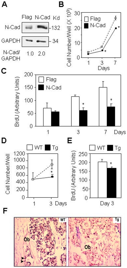
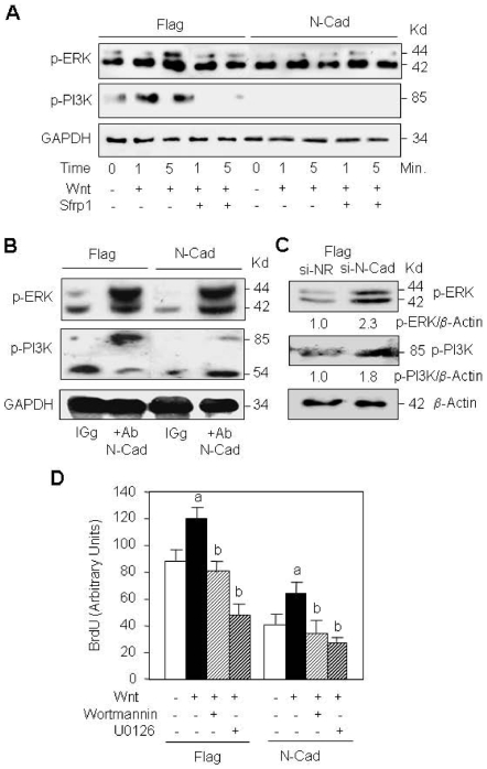
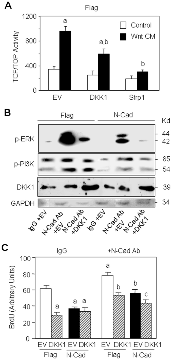
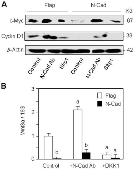
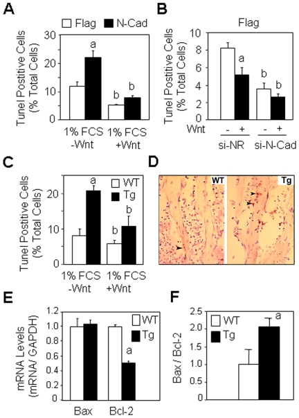
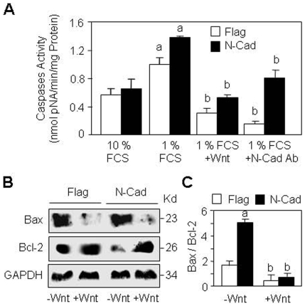
Methods and principal findings: Using murine MC3T3-E1 osteoblastic cells and N-cadherin transgenic mice, we demonstrate that N-cadherin overexpression inhibits cell proliferation in vitro and in vivo. The negative effect of N-cadherin on cell proliferation results from decreased Wnt, ERK and PI3K/Akt signalling and is restored by N-cadherin neutralizing antibody that antagonizes N-cadherin-LRP5 interaction. Inhibition of Wnt signalling using DKK1 or Sfrp1 abolishes the ability of N-cadherin blockade to restore ERK and PI3K signalling and cell proliferation, indicating that the altered cell growth in N-cadherin overexpressing cells is in part secondary to alterations in Wnt signalling. Consistently, we found that N-cadherin overexpression inhibits the expression of Wnt3a ligand and its downstream targets c-myc and cyclin D1, an effect that is partially reversed by N-cadherin blockade. We also show that N-cadherin overexpression decreases osteoblast survival in vitro and in vivo. This negative effect on cell survival results from inhibition of PI3K/Akt signalling and increased Bax/Bcl-2, a mechanism that is rescued by Wnt3a.

Conclusion: The data show that N-cadherin negatively controls osteoblast proliferation and survival via inhibition of autocrine/paracrine Wnt3a ligand expression and attenuation of Wnt, ERK and PI3K/Akt signalling, which provides novel mechanisms by which N-cadherin regulates osteoblast number.


Conflict of interest statement

Competing Interests: The authors have declared that no competing interests exist.

Figures

Figure 1
Figure 1. Enforced expression of N-cadherin decreases cell proliferation in osteoblasts.
(A) MC3T3-E1 osteoblasts stably transfected with N-cadherin (N-Cad) display a 2-fold increase in N-cadherin expression compared to control cells (Flag) as shown by western blot analysis. (B, C) Decreased cell number and replication in N-Cad cells compared to Flag cells. (D, E) Decreased cell number and replication in primary calvarial osteoblasts isolated from N-cadherin transgenic mice (Tg) compared to osteoblasts from wild-type mice (WT). Means are +/− SD. Values that are significantly different are indicated (*, P<0.05 vs Flag or WT cells). (F) Histologial sections of tibias showing decreased cell proliferation in N-Cad Tg mice compared to WT mice, as revealed by Ki67 staining (black nuclei) in bone marrow stromal cells and mature osteoblasts (Ob, arrows) (x250).
Figure 2
Figure 2. N-cadherin reduces cell proliferation via interaction with LRP5 and Wnt signalling.
(A) Immunoprecipitation analysis showing N-cadherin and LRP5 interaction in control (Flag) cells and N-cadherin (N-Cad) overexpressing cells which is blocked by N-cadherin antibody. Cells were treated with blocking N-cadherin antibody (Ab) or control antibody (IgG) for 24 hours, cell lysates were immunoprecipitated (IP) with N-cadherin antibody and analysed by Western-blot (WB) with LRP5 antibody. LRP5, N-cadherin and GAPDH in total proteins were used as loading controls. (B) Treatment with Wnt3a or blocking N-cadherin antibody restores cell proliferation in N-Cad cells. Flag and N-Cad cells were transfected with DKK1 expression vector or empty vector (EV), treated with Wnt3a CM (15%) or N-cadherin antibody for 24 hours and cell replication was determined. Means are +/− SD. Values that are significantly different are indicated (a, P<0.05 vs untreated Flag EV cells; b, P<0.05 vs Flag EV cells treated with N-Cad Ab or Wnt3a; c, P<0.05 vs N-Cad EV cells treated with N-Cad Ab or Wnt3a). (C) N-cadherin silencing increases osteoblast proliferation. Flag cells were transfected with a specific N-cadherin si-RNA or a non relevant si-RNA (si-NR) and treated with Wnt3a CM (15%) for 24 hours and cell replication was determined (a, P<0.05 vs -Wnt si-NR treated cells; b, P<0.05 vs -Wnt si-N-Cad treated cells).
Figure 3
Figure 3. N-cadherin overexpression negatively regulates ERK and PI3K signalling.
(A) Control (Flag) and N-cadherin (N-Cad) overexpressing cells were treated with canonical Wnt3a CM (15%) for 1 or 5 minutes and ERK and PI3K signalling was analysed by Western-blot. GAPDH was used as loading control. (B) N-cadherin blockade restores cell signalling in N-cadherin overexpressing osteoblasts. Flag and N-Cad cells were treated with N-cadherin antibody or control antibody (IgG) for 5 min and ERK and PI3K signalling was analysed by Western-blot. GAPDH was used as loading control. (C) N-cadherin silencing increases ERK and PI3K signalling. Flag cells were transfected with a specific N-cadherin si-RNA or a non relevant si-RNA (si-NR) and phospho-ERk and phospho-PI3K levels determined by western blot analysis were quantified using β-actin as loading control. (D) Treatment with PI3K and MEK inhibitors (Wortmannin and U0126, respectively) abolished cell proliferation induced by Wnt3a CM (15%) in both Flag and N-Cad cells at 24 hours. Means are +/− SD. Values that are significantly different are indicated (a, P<0.05 vs untreated cells; b, P<0.05 vs Wnt3a-treated cells).
Figure 4
Figure 4. The Wnt inhibitor DKK1 abolishes ERK and PI3K signalling restored by N-cadherin blockade.
(A) Control (Flag) cells were transiently transfected with empty vector (EV) or DKK1, or treated with the Wnt antagonist Sfrp1 in the presence or absence of Wnt3a CM and TCF/TOP transcriptional activity was determined. Means are +/− SD. Values that are significantly different are indicated (a, P<0.05 vs EV -Wnt treated cells; b, P<0.05 vs EV Wnt treated cells). (B) Flag and N-Cad cells transiently transfected with empty vector (EV) or DKK1 were treated with the bloking N-cadherin antibody (N-Cad Ab) or control antibody (IgG) for 24 hours and DKK1 levels and ERK and PI3K signalling were analysed by Western-blot. GAPDH was used as loading control. (C) Flag and N-Cad overexpressing cells transfected with empty vector (EV) or DKK1 were treated with the N-cadherin antibody for 24 hours to restore ERK and PI3K signalling or with control antibody (IgG), and cell replication was determined (a, P<0.05 vs EV Flag cells; b, P<0.05 vs N-Cad Ab treated EV Flag cells; c, P<0.05 vs N-Cad Ab treated EV N-Cad cells).
Figure 5
Figure 5. The altered cell proliferation induced by N-cadherin overexpression involves Wnt-responsive genes.
(A) Control (Flag) and N-cadherin (N-Cad) overexpressing cells were treated with the blocking N-cadherin antibody, control antibody (IgG) or the Wnt antagonist Sfrp1 for 24 hours and the levels of the Wnt-responsive proteins c-Myc and cyclin D1 were analysed by Western-blot. β-actin was used as loading control. (B) Flag and N-Cad overexpressing cells were treated with N-cadherin antibody (N-Cad Ab) or IgG, or transiently transfected with the Wnt antagonist DKK1 and Wnt3a mRNA levels were determined by qPCR analysis at 24 hours. Means are +/− SD. Values that are significantly different are indicated (a, P<0.05 vs untreated Flag cells; b, P<0.05 vs corresponding Flag cells).
Figure 6
Figure 6. Forced expression of N-cadherin decreases cell survival.
(A) Control (Flag) and N-cadherin (N-Cad) overexpressing cells cultured in serum deprived (1% FCS) medium were treated with Wnt3a (15% CM) for 24 hours and the number of TUNEL-positive cells was determined. Means are +/− SD (a, P<0.05 vs -Wnt Flag cells; b, P<0.05 vs corresponding Flag cells). (B) N-cadherin silencing decreases osteoblast apoptosis. Flag cells were transfected with N-cadherin si-RNA or a non relevant si-RNA (si-NR) and treated with Wnt3a CM (15%) for 24 hours in serum deprived (1% FCS) medium and cell replication was determined. (a, P<0.05 vs -Wnt si-NR cells; b, P<0.05 vs corresponding Flag cells). (C) Osteoblasts isolated from calvaria in wild type (WT) or N-cadherin transgenic mice (Tg) were cultured in serum deprived (1% FCS) medium and treated with Wnt3a (15% CM) for 24 hours and the number of TUNEL-positive cells was determined (a, P<0.05 vs WT cells; b, P<0.05 vs corresponding WT cells. (D) Histologial sections of tibias showing increased cell apoptosis in Tg mice compared to WT mice, as revealed by TUNEL staining (brown nuclei) in osteoblasts (Ob, arrows) (x250). (E, F) Decreased Bcl-2 mRNA levels and increased Bax/Bcl-2 ratio in tibias of Tg mice compared to WT mice (a, P<0.05 vs WT mice).
Figure 7
Figure 7. Mechanisms by which N-cadherin decreases cell survival.
(A) Control (Flag) and N-Cadherin overexpressing cells (N-Cad) were cultured in survival conditions (10% FCS) or serum deprived (1% FCS) medium, treated with Wnt3a (15% CM), blocking N-cadherin antibody or control antibody (IgG) for 24 hours and effector caspase activity was determined. Means are +/− SD (a, P<0.05 vs corresponding 10% FCS; b, P<0.05 vs corresponding untreated cells. (B) Flag and N-Cad cells were cultured in serum deprived (1% FCS) medium, treated with Wnt3a (15% CM) for 24 hours, and the levels of Bax and Bcl-2 proteins were analysed by Western blot using GAPDH as loading control. (C) Quantification of western blots showing the increased Bax/Bcl-2 ratio in N-Cad cells which was abolished by Wnt3a (a, P<0.05 vs untreated Flag cells; b, P<0.05 vs corresponding untreated cells).
Figure 8
Figure 8. The altered cell survival induced by N-cadherin overexpression is Wnt- and PI3K/Akt-dependent.
(A) Control (Flag) and N-cadherin (N-Cad) overexpressing cells cultured in serum deprived (1% FCS) medium were treated with canonical Wnt3a (15% CM) or the Wnt antagonist Sfrp1 for 1 to 5 minutes and Akt signalling was analysed by Western-blot. GAPDH was used as loading control. (B) Flag and N-Cad overexpressing cells cultured in serum deprived (1% FCS) medium were treated with the blocking N-cadherin antibody, control antibody (IgG) or Sfrp1 and PI3K/Akt signalling was analysed by Western-blot. (C) Flag and N-Cad cells cultured in serum deprived (1% FCS) medium were treated with canonical Wnt3a (15% CM) for 24 hours in the presence or absence of the MEK inhibitor U0126 or the PI3k inhibitor wortmannin, and effector caspase activity was determined. Means are +/− SD. Values that are significantly different are indicated (a, P<0.05 vs untreated Flag cells; b, P<0.05 vs corresponding untreated cells; c, P<0.05 vs corresponding Wnt3a-treated cells). (D) Proposed mechanisms by which N-cadherin acts as a negative regulator of cell proliferation and survival in osteoblasts. N-cadherin interaction with LRP5 (and other proteins: OP) decreases the expression of the autocrine/paracrine Wnt3a ligand and Wnt responsive genes c-Myc and cyclin D1, and causes attenuation of Wnt, ERK and PI3K/Akt signalling, resulting in inhibition of cell proliferation and survival.

Similar articles

Cited by

References

    1. Cadigan KM, Nusse R. Wnt signaling: a common theme in animal development. Genes Dev. 1997;11:3286–3305. - PubMed
    1. Chen S, Guttridge DC, You Z, Zhang Z, Fribley A, et al. Wnt-1 signaling inhibits apoptosis by activating beta-catenin/T cell factor-mediated transcription. J Cell Biol. 2001;152:87–96. - PMC - PubMed
    1. Moon RT, Bowerman B, Boutros M, Perrimon N. The promise and perils of Wnt signaling through beta-catenin. Science. 2002;296:1644–1646. - PubMed
    1. Nusse R, Fuerer C, Ching W, Harnish K, Logan C, et al. Wnt signaling and stem cell control. Cold Spring Harb Symp Quant Biol. 2008;73:59–66. - PubMed
    1. Veeman MT, Axelrod JD, Moon RT. A second canon. Functions and mechanisms of beta-catenin-independent Wnt signaling. Dev Cell. 2003;5:367–377. - PubMed

Publication types

MeSH terms

Substances