Erk1 and Erk2 regulate endothelial cell proliferation and migration during mouse embryonic angiogenesis
- PMID: 20011539
- PMCID: PMC2789384
- DOI: 10.1371/journal.pone.0008283
Erk1 and Erk2 regulate endothelial cell proliferation and migration during mouse embryonic angiogenesis
Abstract
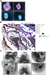
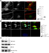
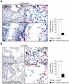
Angiogenesis is a complex process orchestrated by both growth factors and cell adhesion and is initiated by focal degradation of the vascular basement membrane with subsequent migration and proliferation of endothelial cells. The Ras/Raf/MEK/ERK pathway is required for EC function during angiogenesis. Although in vitro studies implicate ERK1 and ERK2 in endothelial cell survival, their precise role in angiogenesis in vivo remains poorly defined. Cre/loxP technology was used to inactivate Erk1 and Erk2 in endothelial cells during murine development, resulting in embryonic lethality due to severely reduced angiogenesis. Deletion of Erk1 and Erk2 in primary endothelial cells resulted in decreased cell proliferation and migration, but not in increased apoptosis. Expression of key cell cycle regulators was diminished in the double knockout cells, and decreased DNA synthesis could be observed in endothelial cells during embryogenesis. Interestingly, both Paxillin and Focal Adhesion Kinase were expressed at lower levels in endothelial cells lacking Erk1 and Erk2 both in vivo and in vitro, leading to defects in the organization of the cytoskeleton and in cell motility. The regulation of Paxillin and Focal Adhesion Kinase expression occurred post-transcriptionally. These results demonstrate that ERK1 and ERK2 coordinate endothelial cell proliferation and migration during angiogenesis.
Conflict of interest statement
Figures


Similar articles
-
ERK2 but not ERK1 mediates HGF-induced motility in non-small cell lung carcinoma cell lines.J Cell Sci. 2013 Jun 1;126(Pt 11):2381-91. doi: 10.1242/jcs.115832. Epub 2013 Apr 2. J Cell Sci. 2013. PMID: 23549785
-
Tissue factor pathway inhibitor (TFPI) interferes with endothelial cell migration by inhibition of both the Erk pathway and focal adhesion proteins.Thromb Haemost. 2008 Mar;99(3):576-85. doi: 10.1160/TH07-10-0623. Thromb Haemost. 2008. PMID: 18327407
-
Genetic demonstration of a redundant role of extracellular signal-regulated kinase 1 (ERK1) and ERK2 mitogen-activated protein kinases in promoting fibroblast proliferation.Mol Cell Biol. 2010 Jun;30(12):2918-32. doi: 10.1128/MCB.00131-10. Epub 2010 Apr 5. Mol Cell Biol. 2010. PMID: 20368360 Free PMC article.
-
Focal adhesion kinase regulation of neovascularization.Microvasc Res. 2012 Jan;83(1):64-70. doi: 10.1016/j.mvr.2011.05.002. Epub 2011 May 14. Microvasc Res. 2012. PMID: 21616084 Free PMC article. Review.
-
ERK1/2 MAP kinases: structure, function, and regulation.Pharmacol Res. 2012 Aug;66(2):105-43. doi: 10.1016/j.phrs.2012.04.005. Epub 2012 Apr 27. Pharmacol Res. 2012. PMID: 22569528 Review.
Cited by
-
Identification of RSK and TTK as Modulators of Blood Vessel Morphogenesis Using an Embryonic Stem Cell-Based Vascular Differentiation Assay.Stem Cell Reports. 2016 Oct 11;7(4):787-801. doi: 10.1016/j.stemcr.2016.08.004. Epub 2016 Sep 8. Stem Cell Reports. 2016. PMID: 27618721 Free PMC article.
-
MAPK Signaling Pathway Plays Different Regulatory Roles in the Effects of Boric Acid on Proliferation, Apoptosis, and Immune Function of Splenic Lymphocytes in Rats.Biol Trace Elem Res. 2024 Jun;202(6):2688-2701. doi: 10.1007/s12011-023-03862-2. Epub 2023 Sep 22. Biol Trace Elem Res. 2024. PMID: 37737440
-
KDM2A promotes lung tumorigenesis by epigenetically enhancing ERK1/2 signaling.J Clin Invest. 2013 Dec;123(12):5231-46. doi: 10.1172/JCI68642. Epub 2013 Nov 8. J Clin Invest. 2013. PMID: 24200691 Free PMC article.
-
miR-132 promotes retinal neovascularization under anoxia and reoxygenation conditions through up-regulating Egr1, ERK2, MMP2, VEGFA and VEGFC expression.Int J Clin Exp Pathol. 2017 Aug 1;10(8):8845-8857. eCollection 2017. Int J Clin Exp Pathol. 2017. PMID: 31966751 Free PMC article.
-
Hepatitis C virus-mediated angiogenesis: molecular mechanisms and therapeutic strategies.World J Gastroenterol. 2014 Nov 14;20(42):15467-75. doi: 10.3748/wjg.v20.i42.15467. World J Gastroenterol. 2014. PMID: 25400432 Free PMC article. Review.
References
-
- Jain RK. Molecular regulation of vessel maturation. Nat Med. 2003;9:685–693. - PubMed
-
- Cheresh DA, Stupack DG. Regulation of angiogenesis: apoptotic cues from the ECM. Oncogene. 2008;27:6285–6298. - PubMed
-
- Liu W, Liu Y, Lowe WL,, Jr, Jr. The role of phosphatidylinositol 3-kinase and the mitogen-activated protein kinases in insulin-like growth factor-I-mediated effects in vascular endothelial cells. Endocrinology. 2001;142:1710–1719. - PubMed
Publication types
MeSH terms
Substances
Grants and funding
LinkOut - more resources
Full Text Sources
Molecular Biology Databases
Research Materials
Miscellaneous

