

Primordial germ cell-like cells differentiated in vitro from skin-derived stem cells
- PMID: 20011593
- PMCID: PMC2788220
- DOI: 10.1371/journal.pone.0008263
Primordial germ cell-like cells differentiated in vitro from skin-derived stem cells
Abstract
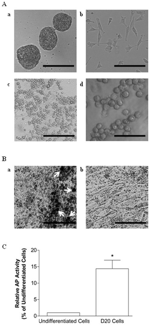
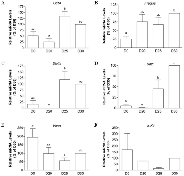
Background: We have previously demonstrated that stem cells isolated from fetal porcine skin have the potential to form oocyte-like cells (OLCs) in vitro. However, primordial germ cells (PGCs), which must also be specified during the stem cell differentiation to give rise to these putative oocytes at more advanced stages of culture, were not systematically characterized. The current study tested the hypothesis that a morphologically distinct population of cells derived from skin stem cells prior to OLC formation corresponds to putative PGCs, which differentiate further into more mature gametes.
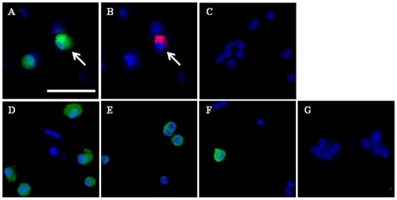
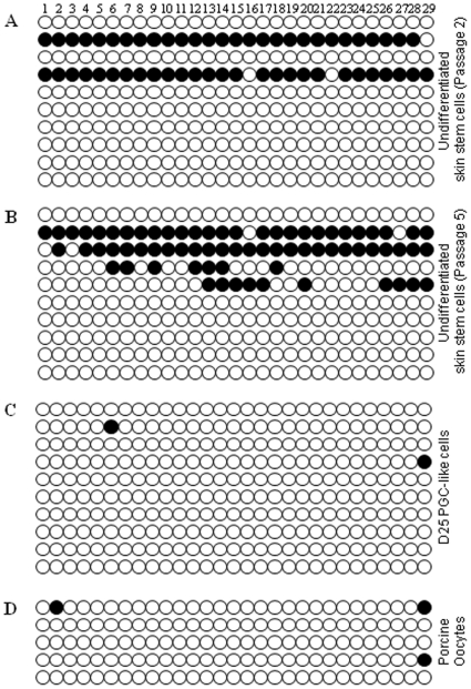
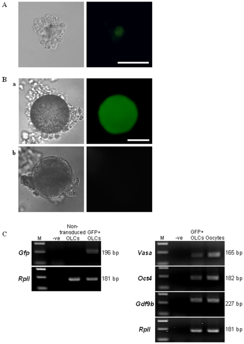
Methodology/principal findings: When induced to differentiate in an appropriate microenvironment, a subpopulation of morphologically distinct cells, some of which are alkaline phosphatase (AP)-positive, also express Oct4, Fragilis, Stella, Dazl, and Vasa, which are markers indicative of germ cell formation. A known differentially methylated region (DMR) within the H19 gene locus, which is demethylated in oocytes after establishment of the maternal imprint, is hypomethylated in PGC-like cells compared to undifferentiated skin-derived stem cells, suggesting that the putative germ cell population undergoes imprint erasure. Additional evidence supporting the germ cell identity of in vitro-generated PGC-like cells is that, when labeled with a Dazl-GFP reporter, these cells further differentiate into GFP-positive OLCs.
Significance: The ability to generate germ cell precursors from somatic stem cells may provide an in vitro model to study some of the unanswered questions surrounding early germ cell formation.
Conflict of interest statement
Figures
Similar articles
-
Analysis of oocyte-like cells differentiated from porcine fetal skin-derived stem cells.Stem Cells Dev. 2011 May;20(5):809-19. doi: 10.1089/scd.2010.0395. Epub 2011 Jan 9. Stem Cells Dev. 2011. PMID: 21054136
-
Stem cells derived from human first-trimester umbilical cord have the potential to differentiate into oocyte-like cells in vitro.Int J Mol Med. 2015 May;35(5):1219-29. doi: 10.3892/ijmm.2015.2132. Epub 2015 Mar 11. Int J Mol Med. 2015. PMID: 25760093 Free PMC article.
-
Deleted in azoospermia-like enhances in vitro derived porcine germ cell formation and meiosis.Stem Cells Dev. 2013 Mar 15;22(6):939-50. doi: 10.1089/scd.2012.0323. Epub 2012 Dec 21. Stem Cells Dev. 2013. PMID: 23259838 Free PMC article.
-
Skin-derived stem cells as a source of primordial germ cell- and oocyte-like cells.Cell Death Dis. 2016 Nov 10;7(11):e2471. doi: 10.1038/cddis.2016.366. Cell Death Dis. 2016. PMID: 27831564 Free PMC article. Review.
-
In vitro differentiation of primordial germ cells and oocyte-like cells from stem cells.Histol Histopathol. 2018 Feb;33(2):121-132. doi: 10.14670/HH-11-917. Epub 2017 Jul 10. Histol Histopathol. 2018. PMID: 28691729 Review.
Cited by
-
Overexpression of Oct4 in porcine ovarian stem/stromal cells enhances differentiation of oocyte-like cells in vitro and ovarian follicular formation in vivo.J Ovarian Res. 2016 Apr 12;9:24. doi: 10.1186/s13048-016-0233-z. J Ovarian Res. 2016. PMID: 27067537 Free PMC article.
-
Human Ovarian Theca-Derived Multipotent Stem Cells Have The Potential to Differentiate into Oocyte-Like Cells In Vitro.Cell J. 2019 Jan;20(4):527-536. doi: 10.22074/cellj.2019.5651. Epub 2018 Aug 1. Cell J. 2019. PMID: 30123999 Free PMC article.
-
Transcriptomic landscape reveals germline potential of porcine skin-derived multipotent dermal fibroblast progenitors.Cell Mol Life Sci. 2023 Jul 22;80(8):224. doi: 10.1007/s00018-023-04869-7. Cell Mol Life Sci. 2023. PMID: 37480481 Free PMC article.
-
Increasing of blastocyst rate and gene expression in co-culture of bovine embryos with adult adipose tissue-derived mesenchymal stem cells.J Assist Reprod Genet. 2016 Oct;33(10):1395-1403. doi: 10.1007/s10815-016-0779-0. Epub 2016 Jul 30. J Assist Reprod Genet. 2016. PMID: 27475633 Free PMC article.
-
The foundational framework of tumors: Gametogenesis, p53, and cancer.Semin Cancer Biol. 2022 Jun;81:193-205. doi: 10.1016/j.semcancer.2021.04.018. Epub 2021 Apr 30. Semin Cancer Biol. 2022. PMID: 33940178 Free PMC article. Review.
References
-
- van den Hurk R, Zhao J. Formation of mammalian oocytes and their growth, differentiation and maturation within ovarian follicles. Theriogenology. 2005;63:1717–1751. - PubMed
-
- Vincent SD, Dunn NR, Sciammas R, Shapiro-Shalef M, Davis MM, et al. The zinc finger transcriptional repressor blimp1/prdm1 is dispensable for early axis formation but is required for specification of primordial germ cells in the mouse. Development. 2005;132:1315–1325. - PubMed
-
- Saitou M, Barton SC, Surani MA. A molecular programme for the specification of germ cell fate in mice. Nature. 2002;418:293–300. - PubMed
-
- Ohinata Y, Payer B, O'Carroll D, Ancelin K, Ono Y, et al. Blimp1 is a critical determinant of the germ cell lineage in mice. Nature. 2005;436:207–213. - PubMed
Publication types
MeSH terms
Substances
Grants and funding
LinkOut - more resources
Full Text Sources
Other Literature Sources
Medical
