. 2009 Dec 14;4(12):e8282.
doi: 10.1371/journal.pone.0008282.

Angiotensin I-converting enzyme mutation (Trp1197Stop) causes a dramatic increase in blood ACE

Affiliations

Angiotensin I-converting enzyme mutation (Trp1197Stop) causes a dramatic increase in blood ACE

Andrew B Nesterovitch et al. PLoS One. .

Abstract

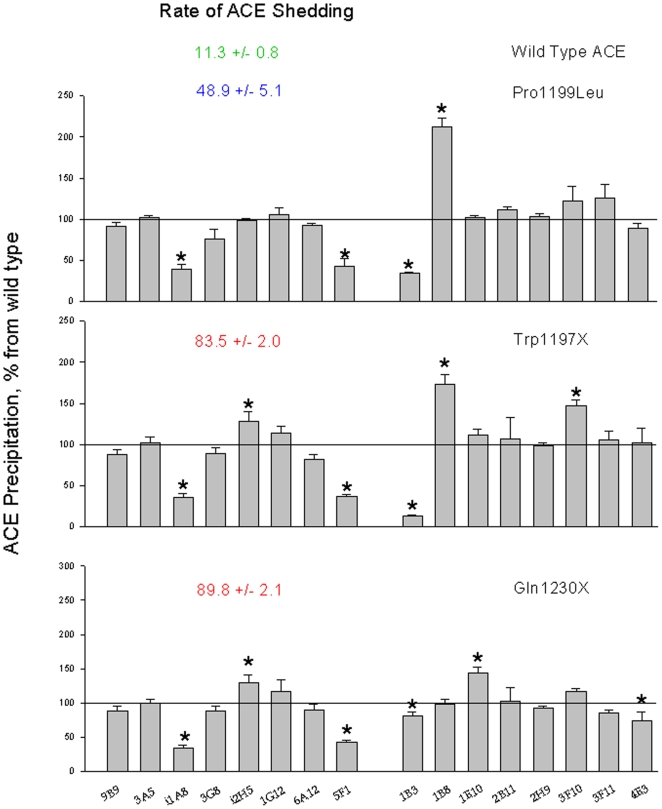
Background: Angiotensin-converting enzyme (ACE) metabolizes many peptides and plays a key role in blood pressure regulation and vascular remodeling. Elevated ACE levels may be associated with an increased risk for different cardiovascular or respiratory diseases, including asthma. Previously, a molecular mechanism underlying a 5-fold familial increase of blood ACE was discovered: Pro1199Leu substitution enhanced the cleavage-secretion process. Carriers of this mutation were Caucasians from Europe (mostly Dutch) or had European roots.

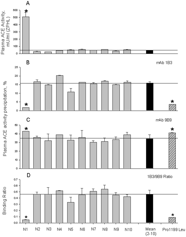
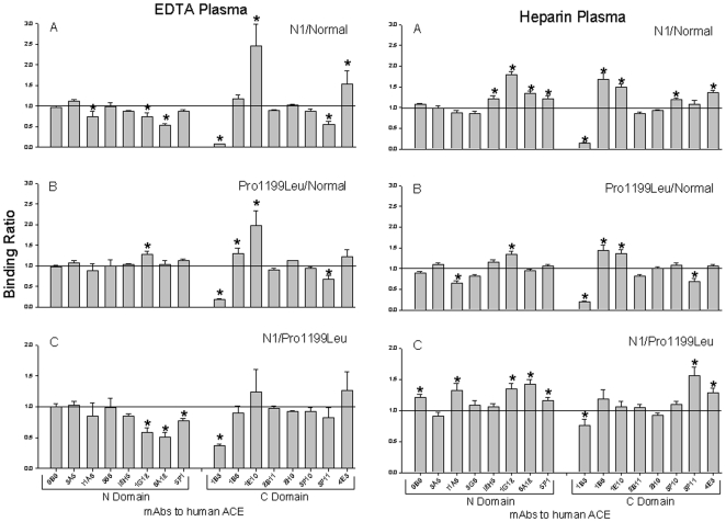
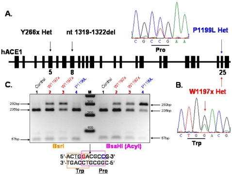
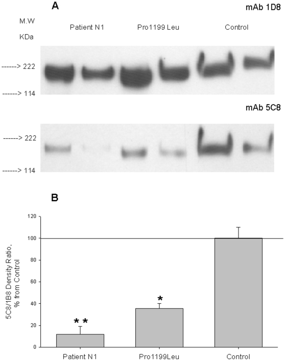
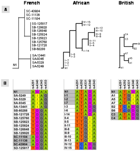
Methodology/principal findings: We have found a family of African-American descent whose affected members' blood ACE level was increased 13-fold over normal. In affected family members, codon TGG coding for Trp1197 was substituted in one allele by TGA (stop codon). As a result, half of ACE expressed in these individuals had a length of 1196 amino acids and lacked a transmembrane anchor. This ACE mutant is not trafficked to the cell membrane and is directly secreted out of cells; this mechanism apparently accounts for the high serum ACE level seen in affected individuals. A haplotype of the mutant ACE allele was determined based on 12 polymorphisms, which may help to identify other carriers of this mutation. Some but not all carriers of this mutation demonstrated airflow obstruction, and some but not all have hypertension.

Conclusions/significance: We have identified a novel Trp1197Stop mutation that results in dramatic elevation of serum ACE. Since blood ACE elevation is often taken as a marker of disease activity (sarcoidosis and Gaucher diseases), it is important for clinicians and medical scientists to be aware of alternative genetic causes of elevated blood ACE that are not apparently linked to disease.


Conflict of interest statement

Competing Interests: The authors have declared that no competing interests exist.

Figures

Figure 1
Figure 1. ACE activity and conformation in the heparinized human plasma.
A. ACE activity in 10 samples of heparinized human plasma from healthy volunteers was quantified using a spectrofluorometric assay with Hip-His-Leu (5 mM) and Z-Phe-His-Leu (2 mM) as substrates. Samples were diluted 1/5 (N2-N10) and 1/80 ( N1). Data expressed as mU/ml (with ZPHL as a substrate) of whole plasma. The results are shown as means + SD of several (3–4) experiments. * - p<0.05 in comparison with mean value for samples N2-N10. B–D. Precipitation of ACE activity from plasma samples by mAbs 1B3 and 9B9. Heparinized plasma samples from of 10 volunteers were equilibrated according to 5 mU/ml of the ACE activity with Hip-His-Leu as a substrate and incubated with a wells on the microtiter plate covered by mAbs 1B3 or 9B9 via goat-anti-mouse IgG; then precipitated ACE activity was quantified by fluorimetric assay - plate precipitation assay , . Data are expressed as a percentage of ACE activity precipitation by mAb 1B3 (B), mAb 9B9 (C) and as their ratio (D). Data are mean ± SD of triplicates. As a positive control sample of pooled plasma sample from several carriers of Pro1199Leu mutation of ACE , was used. * - p<0.05 in comparison with mean value for samples N2-N10.
Figure 2
Figure 2. Conformational fingerprinting of mutant ACEs with a set of mAbs to ACE.
Sixteen monoclonal antibodies were used to precipitate ACE from plasma anticoagulated using EDTA or heparin as indicated. Immunoprecipitated ACE activity is presented as a normalized value (“binding ratio”), to highlight differences in immunoprecipitation pattern (“conformational fingerprint”) among ACE variants from healthy volunteers (“Normal”), from subject N1 (“N1”), or carriers of the Pro1199Leu mutation (“Pro1199Leu”). (A) ACE binding ratios observed for N1 normalized to normal individuals; (B) ACE binding ratios observed for subjects with known Pro1199Leu mutation normalized to normal individuals; (C) ratio of ACE precipitation from subject N1 to that from carriers of Pro1199Leu mutation. Data presented as a mean of 6–8 independent determinations. * p<0.05 indicates ratio shown is significantly different from 1. These ratio for any pair of samples with normal ACE or with any pair of samples with one (Trp1197Stop) or another (Pro1199Leu) identical mutation was around 1.0 with no more than 10% of standard deviation (not shown).
Figure 3
Figure 3. Organization of the identified mutations in ACE gene.
Diagram shows intron-exon structure of human ACE1 gene and localization of the ACE1 mutations. A. Y266x Het mutation in 5th exon and nt1319–1322del TGGA Hom mutation in 8th exon were described in and Pro1199Leu Het mutation in 25th exon – in –. B. Heterozygous mutation Trp1197Stop (W1197x) in 25th exon was revealed by the sequencing of the whole ACE gene (∼24 kb) and confirmed by the restriction analysis of the 292 bp PCR product flanking the part of exon 25 and intron 25 with restriction endonucleases BsrI and BsaHI (AcyI) (C), which was performed with genomic DNA of healthy individual with normal ACE (mother of N1) (1), patient N1 (2) and father of N1 (3) caring mutation Trp1197Stop, and carrier of Pro1199Leu mutation (4). Trp1197Stop (but not Pro1199Leu) mutation abolished restriction site for BsrI.
Figure 4
Figure 4. Family tree of subject N1 with new (Trp1197Stop) mutation in ACE gene.
Individuals with Trp1197Stop mutation are indicated by red color. Genotyped individuals without Trp1197Stop mutation marked by blue color. Individuals found to have high level of serum ACE are marked by upward pointing arrows. Individuals found to have normal level of serum ACE are marked by horizontal arrows. Following abbreviations are used for known clinical diagnoses: HTN – hypertension, DM – diabetes mellitus, HD – heart disease, AFO – airflow obstruction, Asthma – asthma. ACE level in the blood of children (C3–C5) was not tested due to ethical considerations. Among individuals in whom both genotyping and serum ACE determination was performed, there was 100% concordance between presence or absence of the Trp1197Stop mutation and elevated or non-elevated serum ACE levels, respectively.
Figure 5
Figure 5. Conformational fingerprinting of mutated ACE with a set of mAbs to ACE.
Data from the analysis of the precipitation of ACE activity from wild type recombinant human ACE, and recombinant mutant ACEs by 16 mAbs to human ACE were expressed as a ratio of ACE precipitation from Pro1199Leu mutant (A) or Trp1197Stop (B) to that for wild type ACE or ratio of ACE precipitation from Trp1197Stop to that from Pro1199Leu mutation (C). Data presented as a mean of three independent determinations (which were not differ more than 10%) for each ratio. * - p<0.05 in comparison with values for wild type ACE (A and B) or with Pro1199Leu mutant (C).
Figure 6
Figure 6. Western blot analysis of normal and mutant ACEs.
A. ACE purified from plasma of patient N1, pooled plasma from 5 patients with hyper-ACE-emia (carriers of Pro1199Leu mutation) and pooled plasma from 5 healthy individuals with normal blood ACE level were equilibrated to 50 mU/ml of ACE activity (HHL as a substrate), boiled and 40 and 20 µl of each sample run in 4–15% radient gel in reducing conditions. Proteins transferred on PVDF-Plus membrane were revealed with 2 µg/ml of indicated mAb. Molecular weight is shown on the left. B. Revelation of blood ACE presented in panel A was quantified by densitometry of the bands. Data presented as a ratio of density with mAb 5C8 to that with mAb 1D8. Revelation of ACE from the blood of carriers of Pro1199Leu or Thr1197Stop (who contains a mixture of normal and mutated ACEs in their blood) by mAb 5C8 dramatically diminished.
Figure 7
Figure 7. Hierarchical clustering of ACE haplotypes from different ethnic populations.
A. Most parsimonious hierarchical classification tree for ACE haplotypes of individuals from four different ethnic origins and patient N1 is shown. Cladistic analysis was based on 13 informative polymorphisms and revealed 6 levels of divergence. Detailed description of samples is shown only for first two levels; for higher divergence only number of samples is indicated in parentheses. The size of boxes correlates with number of samples within the clade. Human populations were CEU, Caucasians; JPT, Japan; YRI, Africans; CHB, Chinese; and N1, patient with Trp1197Stop mutation. N1 patient is similar to Nigerians YRI-19211 and YRI-18517, and one Chinese CHB-18577. B. ACE genotypes and haplotypes for patient N1 and individuals from levels 1, 2 and 3 are shown. Positions highlighted in green were conservative in all shown individuals. Polymorphic nucleotides in N1 are highlighted in red, and show in alternative colors (yellow vs. blue) in other clades/individuals. Some positions were C/T or A/G heterozygous; or genotyping data were missing (N/N). SNPs (“rs” with number), position of the I/D polymorphism, and the position of W1197X mutation in N1 are shown on the top of the table. Linkage disequilibrium plot for Nigerian population is presented on the bottom (source: International HapMap project at http://www.hapmap.org).
Figure 8
Figure 8. Clustering of the patient N1 haplotype into ACE clades.
A. Clustering of the patient N1 haplotype along clades identified in French , British and African-originated populations. B. Detailed haplotypes of identified clades and N1 are presented. N1 carrier of the W1197X mutation perfectly matched with Africa-originated clade I (sub-clades 1, 2, and 7) and was consistently associated with clades represented high ACE plasma level (see text).

Similar articles

Cited by

References

    1. Ehlers MR, Riordan JF. Angiotensin-converting enzyme: new concepts concerning its biological role. Biochemistry. 1989;8:5311–5318. - PubMed
    1. Corvol P, Eyries M, Soubrier F. Peptidyl-dipeptidase A/Angiotensin I-converting enzyme. In: Barret A, Rawlings N, Woessner J, editors. Handbook of Proteolytic Enzymes. New York: Elsevier Academic Press; 2004. pp. 332–349.
    1. Bernstein KE, Xiao HD, Frenzel K, Li P, Shen XZ, et al. Six truisms concerning ACE and the renin-angiotensin system educed from the genetic analysis of mice. Circ Res. 2005;96:1135–1144. - PubMed
    1. Caldwell PR, Seegal BC, Hsu KC, Das H, Soffer RL. Angiotensin-converting enztme: vascular endothelial localization. Science. 1976;191:1050–1051. - PubMed
    1. Defendini R, Zimmerman EA, Weare JA, Alhenc-Gelas F, Erdos EG. Angiotensin-converting enzyme in epithelial and neuroepithelial cells. Neuroendocrinology. 1983;37:32–40. - PubMed

Publication types

MeSH terms