. 2009 Sep 1;7(4):358-83.
doi: 10.2203/dose-response.09-027.Hamblin.

Biphasic dose response in low level light therapy

Affiliations

Biphasic dose response in low level light therapy

Ying-Ying Huang et al. Dose Response. .

Abstract

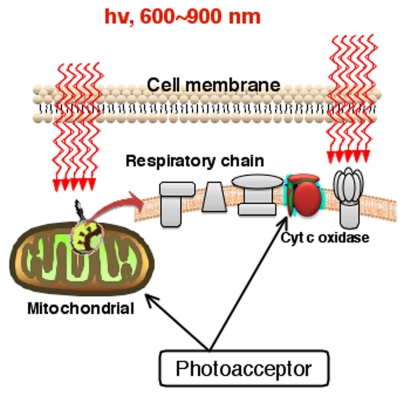
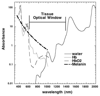
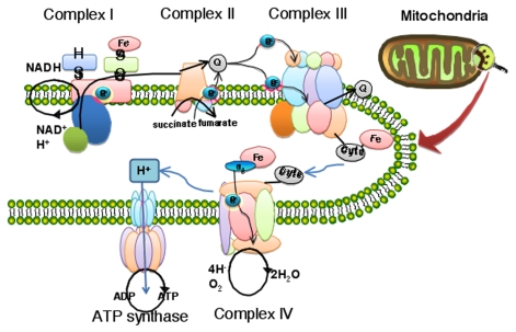
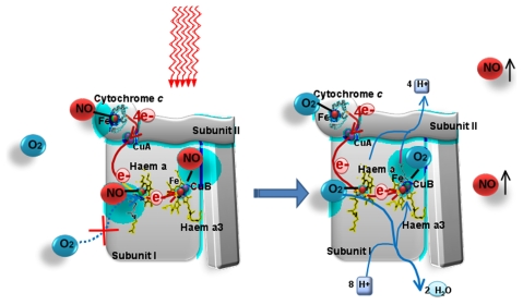
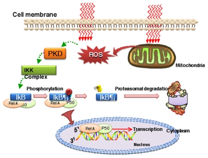
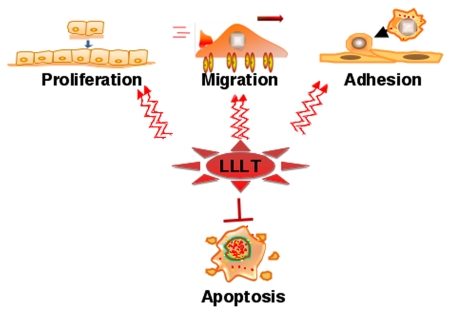
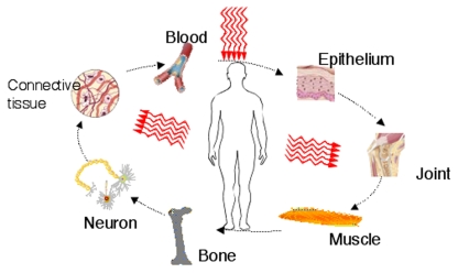
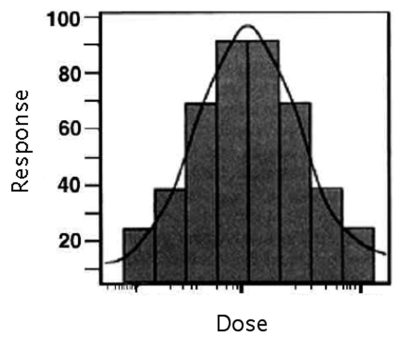
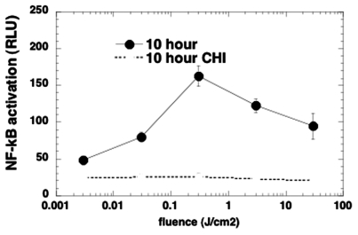
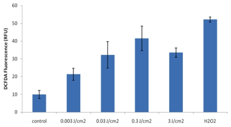
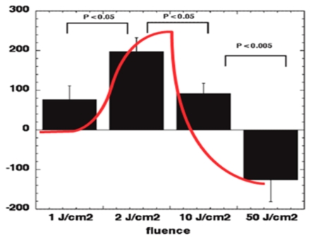
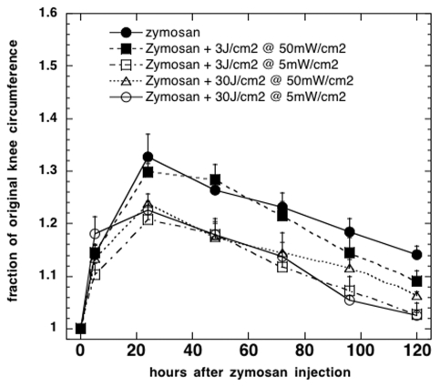
The use of low levels of visible or near infrared light for reducing pain, inflammation and edema, promoting healing of wounds, deeper tissues and nerves, and preventing cell death and tissue damage has been known for over forty years since the invention of lasers. Despite many reports of positive findings from experiments conducted in vitro, in animal models and in randomized controlled clinical trials, LLLT remains controversial in mainstream medicine. The biochemical mechanisms underlying the positive effects are incompletely understood, and the complexity of rationally choosing amongst a large number of illumination parameters such as wavelength, fluence, power density, pulse structure and treatment timing has led to the publication of a number of negative studies as well as many positive ones. A biphasic dose response has been frequently observed where low levels of light have a much better effect on stimulating and repairing tissues than higher levels of light. The so-called Arndt-Schulz curve is frequently used to describe this biphasic dose response. This review will cover the molecular and cellular mechanisms in LLLT, and describe some of our recent results in vitro and in vivo that provide scientific explanations for this biphasic dose response.


Figures

FIGURE 1
FIGURE 1
Schematic diagram showing the absorption of red and NIR light by specific cellular chromophores or photoacceptors localized in the mitochondrial respiratory chain
FIGURE 2
FIGURE 2
Absorption spectra of the main chromophores in living tissue on a log scale showing the optical window where visible and NIR light can penetrate deepest into tissue.
FIGURE 3
FIGURE 3
Mitochondrial respiratory chain consisting of contains five complexes of integral membrane proteins: NADH dehydrogenase (Complex I), succinate dehydrogenase (Complex II), cytochrome c reductase (Complex III), cytochrome c oxidase (Complex IV), and ATP synthase (ComplexV).
FIGURE 4
FIGURE 4
When NO is released from its binding to heme iron and copper centers in cytochrome c oxidase by the action of light, oxygen is allowed to rebind to these sites and respiration is restored to its former level leading to increased ATP synthesis.
FIGURE 5
FIGURE 5
Reactive oxygen species (ROS) formed as a result of LLLT effects in mitochondria may activate the redox-sensitive transcription factor NF-κB (relA-p50) via protein kinase D (PKD).
FIGURE 6
FIGURE 6
The downstream cellular effects of LLLT signaling include increases in cell proliferation, migration and adhesion molecules. Cell survival is increased and cell death reduced by expression of proteins that inhibit apoptosis.
FIGURE 7
FIGURE 7
Beneficial tissue effects of LLLT can include almost all the tissues and organs of the body.
FIGURE 8
FIGURE 8
Idealized biphasic dose response curve (often termed Arndt-Schulz curve) typically reported in LLLT studies.
FIGURE 9
FIGURE 9
Biphasic dose response of NF-κB activation (measured by bioluminescence reporter assay) in mouse embryonic fibroblasts 10 hours after different fluences of 810-nm laser light. CHI is control where all protein synthesis has been inhibited.
FIGURE 10
FIGURE 10
Biphasic dose response in generation of ROS as detected by fluorescence probe under same conditions as Fig 9 but measured at 5 minutes post-irradiation.
FIGURE 11
FIGURE 11
Biphasic dose response in measured difference in integrated area under the curve of time course of wound size compared to no treatment control, with a clear maximum seen at 2 J/cm2, and the high dose of 50 J/cm2 gave a worsening of the wound healing time curve.
FIGURE 12
FIGURE 12
Dose response of illumination time found in a study of 810-nm laser to treat zymosan-induced arthritis in rats. Integrated curves of knee circumference versus time were compared. Three LLLT regimens were equally successful where the illumination time was either 100 minutes or 10 minutes, but the ineffective regimen only had a 1 minute illumination time.

Similar articles

Cited by

References

    1. Ad N, Oron U. Impact of low level laser irradiation on infarct size in the rat following myocardial infarction. Int J Cardiol. 2001;80:109–16. - PubMed
    1. Agaiby AD, Ghali LR, Wilson R, Dyson M. Laser modulation of angiogenic factor production by T-lymphocytes. Lasers Surg Med. 2000;26:357–63. - PubMed
    1. Aimbire F, Albertini R, Pacheco MT, Castro-Faria-Neto HC, Leonardo PS, Iversen VV, Lopes-Martins RA, Bjordal JM. Low-level laser therapy induces dose-dependent reduction of TNFalpha levels in acute inflammation. Photomed Laser Surg. 2006;24:33–7. - PubMed
    1. al-Watban FA, Andres BL. The effect of He-Ne laser (632.8 nm) and Solcoseryl in vitro. Lasers Med Sci. 2001;16:267–75. - PubMed
    1. Alexandratou E, Yova D, Handris P, Kletsas D, Loukas S. Human fibroblast alterations induced by low power laser irradiation at the single cell level using confocal microscopy. Photochem Photobiol Sci. 2002;1:547–52. - PubMed

LinkOut - more resources