. 2009 Dec 8;4(12):e8198.
doi: 10.1371/journal.pone.0008198.

ActRIIA and BMPRII Type II BMP receptor subunits selectively required for Smad4-independent BMP7-evoked chemotaxis

Affiliations

ActRIIA and BMPRII Type II BMP receptor subunits selectively required for Smad4-independent BMP7-evoked chemotaxis

Jeanette C Perron et al. PLoS One. .

Abstract

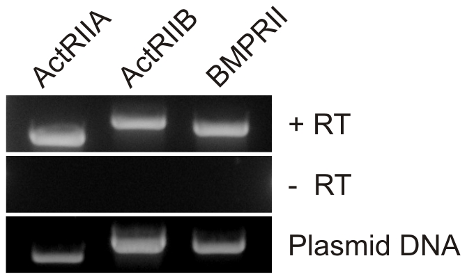
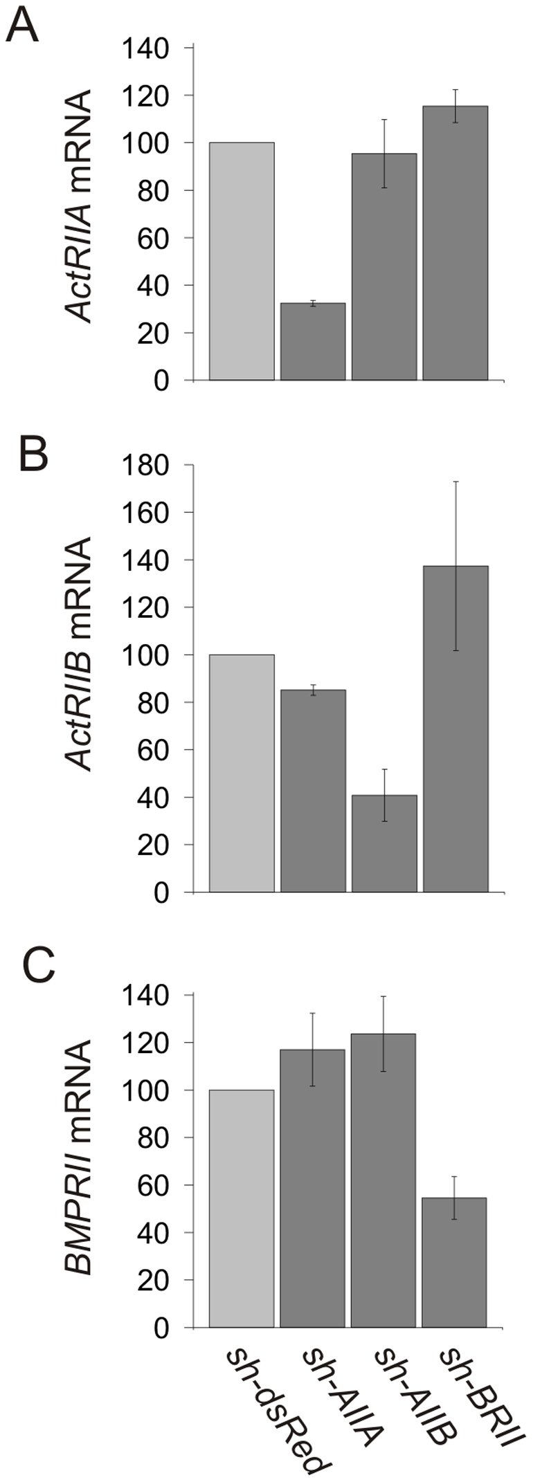
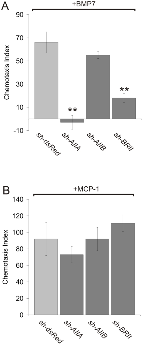
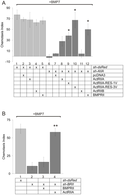
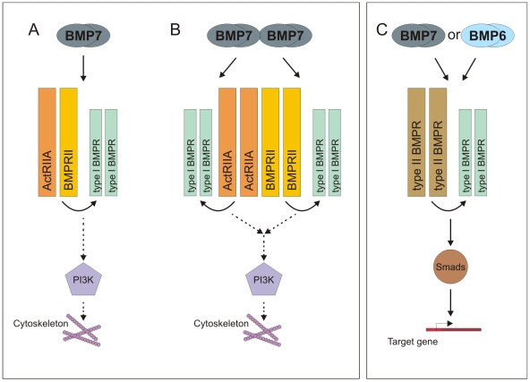
Bone morphogenetic protein (BMP)-evoked reorientation and chemotaxis of cells occurs with rapid onset and involves events local to the cell membrane. The signaling pathways underlying these rapid processes likely diverge from those mediating classical transcriptional responses to BMPs but it remains unclear how BMP receptors are utilized to generate distinct intracellular mechanisms. We show that BMP7-evoked chemotaxis of monocytic cells depends on the activity of canonical type II BMP receptors. Although the three canonical type II BMP receptors are expressed in monocytic cells, inhibition of receptor subunit expression by RNAi reveals that ActRIIA and BMPRII, but not ActRIIB, are each essential for BMP7-evoked chemotaxis but not required individually for BMP-mediated induction. Furthermore, the chemotactic response to BMP7 does not involve canonical Smad4-dependent signaling but acts through PI3K-dependent signaling, illustrating selective activation of distinct intracellular events through differential engagement of receptors. We suggest a model of a BMP receptor complex in which the coordinated activity of ActRIIA and BMPRII receptor subunits selectively mediates the chemotactic response to BMP7.


Conflict of interest statement

Competing Interests: The authors have declared that no competing interests exist.

Figures

Figure 1
Figure 1. Chemotaxis of monocytic cells in response to a panel of BMPs.
A: WEHI 274.1 cell migration through transwell chamber filters in response to 10 pg/ml BMP7, BMP2, BMP6 or GDF7 in the lower chamber. For comparison, cells were also stimulated with MCP-1 (100 ng/ml). The chemotaxis index (CI) is shown as mean +/− SEM. MCP-1 (CI = 139+/−45, n = 4); BMP7 (CI = 93+/−38, n = 5); BMP2 (CI = 116+/−40, n = 4); BMP6 (CI = −23+/−6, n = 3); GDF7 (CI = 10+/−10, n = 3). B: Dose response curves for chemotaxis of WEHI 274.1 cells in response to BMP7, BMP6 and GDF7 (n≥3 for each protein at each concentration). Migration in the presence of BMP7 differs significantly from migration in the presence of BMP6 or of GDF7; (*) p<0.05; (**) p<0.01; (***) p<0.002 (Student's t test).
Figure 2
Figure 2. Type II BMP receptor mRNA expression in WEHI 274.1 cells.
cDNA from WEHI 274.1 cells transcribed from total RNA in the presence (top, +RT) and absence (middle, -RT) of reverse transcriptase and amplified with BMP receptor subunit-specific primers. Plasmids encoding type II BMP receptor cDNAs (bottom, Plasmid DNA) were used as a positive control for each primer set. The sizes of the expected reaction products are 702 bp for ActRIIA, 928 bp for ActRIIB, and 813 bp for BMPRII.
Figure 3
Figure 3. Knockdown of type II BMP receptors in WEHI 274.1 cells by receptor-targeted shRNAs.
Real-time QPCR analysis of dsRedΔ−, ActRIIAΔ−, ActRIIBΔ− and BMPRIIΔWEHI cells. Results are expressed as the percent of control (mean +/− SEM) for each condition relative to BMP receptor mRNA levels in dsRedΔWEHI (control) cells (n = 2). A: ActRIIA mRNA expression in the presence of sh-AIIA (32%+/−1%), sh-AIIB (95%+/−14%) or sh-BRII (115%+/−7%). B: ActRIIB mRNA expression in the presence of sh-AIIA (85%+/−2%), sh-AIIB (41%+/−11%) or sh-BRII (137%+/−36%). C: BMPRII mRNA expression in the presence of sh-AIIA (117%+/−15%), sh-AIIB (124%+/−16%) or sh-BRII (55%+/−9%). Individual type II BMP receptor shRNAs modulated target mRNAs selectively, but had no significant effect on the expression level of non-target BMP receptor mRNA.
Figure 4
Figure 4. BMP7-stimulated WEHI 274.1 cell chemotaxis following type II BMP receptor knockdown.
Chemotaxis of dsRedΔ−, ActRIIAΔ−, ActRIIBΔ− and BMPRIIΔWEHI cells (mean +/− SEM) in response to BMP7 (10 pg/ml, A) or MCP-1 (100 ng/ml, B). In A, dsRedΔWEHI (CI = 66+/−9; n = 8), ActRIIAΔWEHI (CI = −3+/−6; n = 3), ActRIIBΔWEHI (CI = 55+/−3; n = 4) and BMPRIIΔWEHI cells (CI = 18+/−4; n = 7). n = 3 for each condition in B. BMP7-stimulated chemotaxis following knockdown with either sh-AIIA or sh-BRII differs significantly from that in dsRedΔWEHI (control) cells; (**) p<0.0001 (Student's t test).
Figure 5
Figure 5. Rescue of shRNA-mediated inhibition of BMP7-stimulated chemotaxis by ActRIIA and BMPRII but not ActRIIB.
A: Chemotaxis in response to 10 pg/ml BMP7 (mean +/− SEM) of dsRedΔWEHI cells (Lanes 1–5): alone (CI = 77+/−9, n = 3) or co-expressing pcDNA3 (CI = 69+/−4, n = 2), ActRIIA (CI = 68+/−14, n = 3), ActRIIB (CI = 63+/−5, n = 2) or BMPRII (CI = 66+/−6, n = 2). These are compared with ActRIIAΔWEHI cells (Lane 6–12): alone (CI = −3+/−6, n = 3) or co-expressing pcDNA3 (CI = 5+/−1, n = 3), wild-type ActRIIA (CI = 28+/−9, n = 3), ActRIIA-RES-1V (CI = 38+/−6, n = 3), ActRIIA-RES-3V (CI = 67+/−14, n = 3), ActRIIB (CI = 5+/−8, n = 3) or BMPRII (CI = 50+/−10, n = 3). B: Chemotaxis in response to 10 pg/ml BMP7 (mean +/− SEM) of dsRedΔWEHI (CI = 63+/−7) and BMPRIIΔWEHI (CI = 11+/−5) cells (Lanes 1 and 2) compared with BMPRIIΔWEHI cells co-expressing wild-type BMPRII (CI = 16+/−7) or ActRIIA (CI = 58+/−5) (Lanes 3 and 4). n = 3 for each condition. BMP7-evoked chemotaxis in ActRIIAΔWEHI cells expressing ActRIIA-RES-1V, ActRIIA-RES-3V or BMPRII differs significantly from chemotaxis in ActRIIAΔWEHI cells alone; (*) p<0.02 (Student's t test). BMP7-evoked chemotaxis in BMPRIIΔWEHI cells expressing wild-type ActRIIA differs significantly from chemotaxis in BMPRIIΔWEHI cells alone; (**) p<0.005 (Student's t test).
Figure 6
Figure 6. Knockdown of Smad4 inhibits BMP7-evoked Id1 induction but not BMP7-evoked chemotaxis.
A: Western blot analysis of lysates of dsRedΔC2C12 and Smad4ΔC2C12 cells, probed for expression of Smad4 and GFP (loading control) and showing 30% knockdown of endogenous Smad4 (mean +/− SEM, n = 3). B: Northern analysis of mouse Id1 expression in control and 150 ng/ml BMP7-stimulated dsRedΔC2C12 and Smad4ΔC2C12 cells (Actin expression used as loading control). Smad4ΔC2C12 cells show a 46% decrease in BMP7-evoked Id1 induction (mean +/− SEM, n = 4). C: Western blots of lysates from dsRedΔWEHI and Smad4ΔWEHI cells, probed for expression of Smad4 and GFP (loading control), demonstrate efficient knockdown of endogenous Smad4 by sh-Smad4 (84% reduction, mean +/− SEM, n = 3) in WEHI 274.1 cells. D: dsRedΔWEHI and Smad4ΔWEHI cell chemotaxis in response to 10 pg/ml BMP7 did not differ (mean +/− SEM, n = 3). dsRedΔWEHI (CI = 159+/−33) v. Smad4ΔWEHI (CI = 140+/−41); p = 0.745 (Student's t test).
Figure 7
Figure 7. PI3K activity is stimulated by BMP7 and required for BMP7-evoked chemotaxis.
A: Western blots of whole cell lysates of WEHI 274.1 cells incubated with or without 50 ng/ml BMP7 were probed with a phospho-specific α-Akt(S) antibody. Measurement of total Akt and GAPDH provided loading controls. Densitometric measurement (mean+/−SEM) shows an increase in response to BMP7 (70% over control n = 2). B: Western blots of whole cell lysates of control and 50 µM LY-treated WEHI 274.1 cells incubated with or without 50 ng/ml BMP7 were probed with a phospho-specific α-Smad1/5/8 antibody. Measurement of total Smad and GAPDH provided loading controls. Inhibition of PI3K activity did not affect BMP7-evoked stimulation of R-Smad phosphorylation. C: Chemotaxis (mean +/− SEM) of WEHI 274.1 cells in response to 10 pg/ml BMP7 was significantly reduced following 50 µM LY294002 (LY) treatment (85% reduction). BMP7-LY (CI = 86+/−15, n = 3) v. BMP7+LY (CI = 13+/−3.5, n = 3), p = 0.0086 (Student's t test). Treatment with LY alone had no effect on WEHI 274.1 cell movement under control conditions (LY alone: CI = 3.5+/−2.5, n = 2; J. C. Perron and J. Dodd, unpublished).
Figure 8
Figure 8. Model of differential receptor subunit recruitment and signaling underlying BMP-evoked chemotaxis and trophic actions.
We have demonstrated that ActRIIA and BMPRII receptor subunits are both selectively required for BMP7-evoked chemotaxis in monocytic cells. How do the individual receptor subunits contribute to this activity? Do they work together in a common complex? A,B: Our data support a model in which BMP7 engages both ActRIIA and BMPRII subunits either as part of a heteromeric type II receptor pair (A) or as homodimers, possibly forming unique receptor complexes combining pairs of each of the type II receptor subunits (B). The identity of the type I receptors that contribute to this complex is not known, although recent evidence implicates BMPRIB in BMP-dependent axon guidance , . Engagement of ActRIIA and BMPRII subunits by chemotropic BMPs activates a signaling pathway leading to PI3K activity and cytoskeletal reorganization that is independent of Smad4-mediated nuclear signaling. C: In contrast, BMP6 does not stimulate chemotaxis, presumably being unable to recruit or activate the ActRIIA:BMPRII complex necessary for chemotropic signaling. In the same cell, however, BMP7 and BMP6 share the ability to activate Smad-dependent transcriptional pathways, through receptor mechanisms that do not depend selectively on either ActRIIA or BMPRII.

Similar articles

Cited by

References

    1. Sanchez-Camacho C, Rodriguez J, Ruiz JM, Trousse F, Bovolenta P. Morphogens as growth cone signalling molecules. Brain Res Brain Res Rev. 2005;49:242–252. - PubMed
    1. Charron F, Tessier-Lavigne M. Novel brain wiring functions for classical morphogens: a role as graded positional cues in axon guidance. Development. 2005;132:2251–2262. - PubMed
    1. Liu A, Niswander LA. Bone morphogenetic protein signalling and vertebrate nervous system development. Nat Rev Neurosci. 2005;6:945–954. - PubMed
    1. Derynck R, Zhang YE. Smad-dependent and Smad-independent pathways in TGF-beta family signalling. Nature. 2003;425:577–584. - PubMed
    1. Shi Y, Massague J. Mechanisms of TGF-beta signaling from cell membrane to the nucleus. Cell. 2003;113:685–700. - PubMed

Publication types

MeSH terms