

Dynamic kine magnetic resonance imaging in whiplash patients and in age- and sex-matched controls
- PMID: 20011712
- PMCID: PMC2807769
- DOI: 10.1155/2009/369612
Dynamic kine magnetic resonance imaging in whiplash patients and in age- and sex-matched controls
Abstract
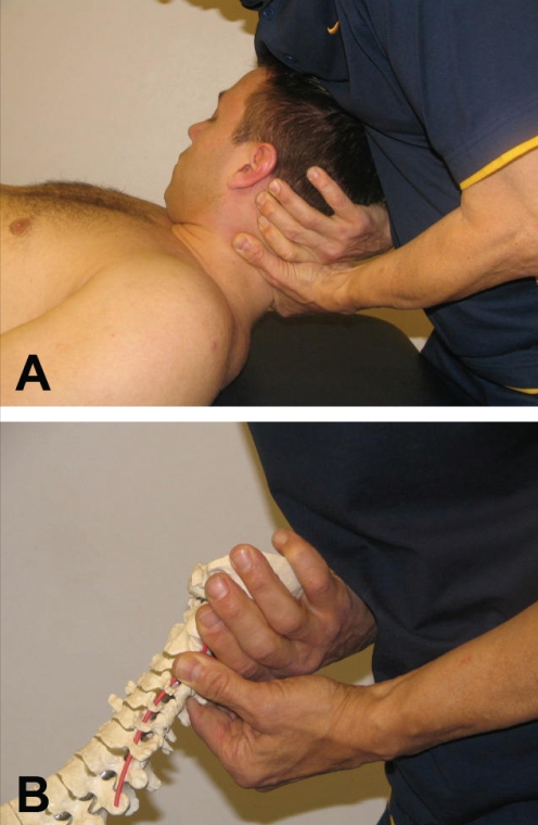
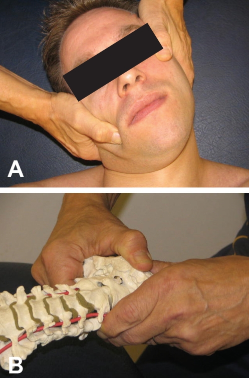
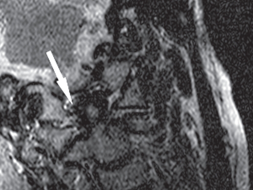
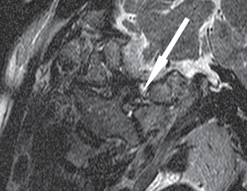
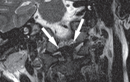
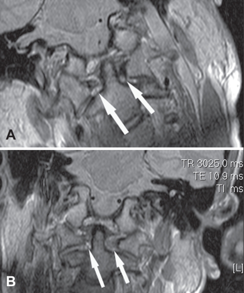
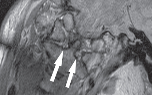
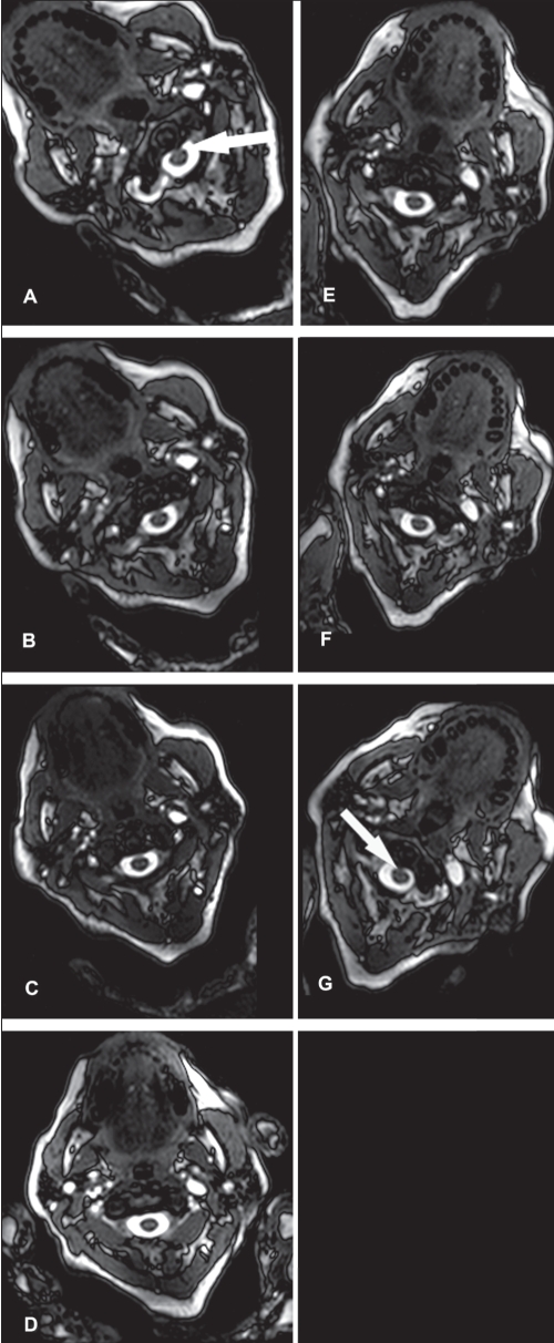
The multitude of symptoms following a whiplash injury has given rise to much discussion because of the lack of objective radiological findings. The ligaments that stabilize the upper cervical spine can be injured. Dynamic kine magnetic resonance imaging (dMRI) may reveal the pathological motion patterns caused by injury to these ligaments. To compare the findings and motion patterns in the upper cervical spine, 25 whiplash trauma patients with longstanding pain, limb symptoms and loss of balance indicating a problem at the level of C0-C2, as well as matched healthy controls were imaged using dMRI. Imaging was performed with an Intera 1.5 T (Philips Healthcare, USA) magnet. A physiotherapist performed the bending and rotation of the upper cervical spine for the subjects to ensure that the movements were limited to the C0-C2 level. An oblique coronal T2- and proton density-weighted sequence and a balanced fast field echo axial sequence were used. The movements between C0-C2 and the signal from the alar ligaments were analyzed. Contact of the transverse ligament and the medulla in rotation was seen in two patients. The signal from the alar ligaments was abnormal in 92% of the patients and in 24% of the control subjects (P<0.0001). Abnormal movements at the level of C1-C2 were more common in patients than in controls (56% versus 20%, P=0.028). Whiplash patients with longstanding symptoms had both more abnormal signals from the alar ligaments and more abnormal movements on dMRI at the C0-C2 level than controls.
La multitude de symptômes après un coup de fouet cervical a suscité de nombreuses discussions en raison de l’absence de résultats radiologiques objectifs. Les ligaments qui peuvent stabiliser la colonne cervicale supérieure peuvent subir un traumatisme. La ciné-imagerie par résonance magnétique dynamique (IRMd) peut révéler le profil de mouvements pathologiques provoqué par le traumatisme de ces ligaments. Pour comparer les observations et le profil de mouvements de la colonne cervicale supérieure, 25 patients ayant subi un traumatisme par coup de fouet cervical ayant des douleurs, des symptômes dans les membres et une perte d’équilibre de longue date et faisant état d’un problème aux vertèbres C0–C2, de même que des sujets témoins appariés en santé, ont subi une IRMd. On a procédé à cette imagerie au moyen d’un aimant Intera 1.5 T (Philips Healthcare, États-Unis). Un physiothérapeute a procédé au fléchissement et à la rotation de la colonne cervicale supérieure chez les sujets pour s’assurer que les mouvements se limitaient aux vertèbres C0–C2. On a utilisé une image coronaire oblique par séquence pondérée T2 et de densité des protons (T2pd) et par séquence axiale équlibrée écho de gradient (FFE). On a analysé le mouvement entre les vertèbres C0–C2 et le signal des ligaments auxiliaires. On a observé un contact entre le ligament transverse et la région médullaire en rotation chez deux patients. Le signal des ligaments auxiliaires était anormal chez 92 % des patients et chez 24 % des sujets témoins (P<0,0001). Les mouvements anormaux au niveau des vertèbres C0–C2 étaient plus courants chez les patients que chez les sujets témoins (56 % par rapport à 20 %, P=0,028). Les patients ayant subi un coup de fouet cervical qui avaient des symptômes de longue date présentaient à la fois plus de signaux anormaux des ligaments auxiliaires et plus de mouvements anormaux au niveau des vertèbres C0–C2 à l’IRMd que les sujets témoins.
Figures
References
-
- Spitzer WO, Skovron MI, Salmi LR, et al. Scientific monograph of the Quebec Task Force on Whiplash Associated Disorders: Redefining whiplash and its management. Spine. 1995;20:1S–73S. - PubMed
-
- Barnsley L, Lord S, Bogduk N. Whiplash injury. Pain. 1994;58:283–307. - PubMed
-
- Barnsley L, Lord SM, Wallis BJ, Bogduk N. The prevalence of chronic cervical zygapophysial joint pain after whiplash. Spine. 1995;20:20–5. - PubMed
-
- Dvorak J, Panjabi MM. Functional anatomy of the alar ligaments. Spine. 1987;12:183–91. - PubMed
Publication types
MeSH terms
LinkOut - more resources
Full Text Sources
Medical
Miscellaneous
