. 2010 Sep;39(9):837-57.
doi: 10.1007/s00256-009-0842-3. Epub 2009 Dec 10.

Ulnar-sided wrist pain. II. Clinical imaging and treatment

Affiliations

Ulnar-sided wrist pain. II. Clinical imaging and treatment

Atsuya Watanabe et al. Skeletal Radiol. 2010 Sep.

Abstract

Pain at the ulnar aspect of the wrist is a diagnostic challenge for hand surgeons and radiologists due to the small and complex anatomical structures involved. In this article, imaging modalities including radiography, arthrography, ultrasound (US), computed tomography (CT), CT arthrography, magnetic resonance (MR) imaging, and MR arthrography are compared with regard to differential diagnosis. Clinical imaging findings are reviewed for a more comprehensive understanding of this disorder. Treatments for the common diseases that cause the ulnar-sided wrist pain including extensor carpi ulnaris (ECU) tendonitis, flexor carpi ulnaris (FCU) tendonitis, pisotriquetral arthritis, triangular fibrocartilage complex (TFCC) lesions, ulnar impaction, lunotriquetral (LT) instability, and distal radioulnar joint (DRUJ) instability are reviewed.


Figures
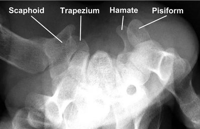
Fig. 1
Fig. 1
Carpal tunnel view of the wrist (radiograph). The carpal tunnel is bounded on the ulnar side by the hook of the hamate and on the radial side by the distal end of the scaphoid. The pisiform is ulnar to the hamate and the volar ridge of the trapezium is radial to the scaphoid
Fig. 2
Fig. 2
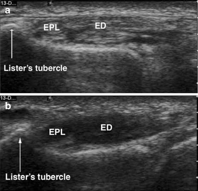
Transverse ultrasound of the extensor surface of the wrist show the extensor digitorum (ED) and extensor pollicis longus (EPL) tendons, a without artifact on the image obtained with the probe held exactly perpendicular to the tendons, b but with a significant loss of echogenicity on the image obtained with the probe held at an oblique angle to the tendons. (Reproduced with permission from [151])
Fig. 3
Fig. 3
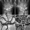
High-resolution wrist MRI with a 2-inch coil at 3.0 T. a Two-dimensional (2D) fast spin echo (TR/TE = 18,00/11.6, field of view 6 cm, matrix 192 × 256, slice thickness 1 mm), b 2D gradient echo (GRE) (TR/TE/FA = 500/15/40, field of view 6 cm, matrix 192 × 256, slice thickness 1 mm), and c 3D GRE TR/TE/flip angle = 31/9/10, field of view 6 cm, matrix 192 × 256, slice thickness 1 mm) demonstrate a small central perforation of the triangular fibrocartilage complex (TFCC)
Fig. 4
Fig. 4
a Coronal fat-suppressed T1-weighted MR arthrographic image (TR/TE = 650/15), b sagittal proton density-weighted image (TR/TE = 2,000/32) show an articular disc tear of the TFCC (arrow) with contrast material within the distal radioulnar joint (arrowhead). Images are suggestive of a peripheral TFCC tear with mild extravasation of the contrast agent (curved arrow)
Fig. 5
Fig. 5
a Axial and b reformatted sagittal CT of the wrist in a patient with hamate fracture (arrows)
Fig. 6
Fig. 6
Kienböck’s disease. Radiographs of a the affected side (arrow) and b the non-affected side. c Coronal proton-density-weighted (TR/TE = 1,582/15) and d coronal GRE (TR/TE/flip angle = 379/12/40) MR imaging of the wrist in the same patient with Kienböck’s disease demonstrate mixed bone marrow edema and sclerosis with slightly collapsed lunate (arrows)
Fig. 7
Fig. 7
High-resolution MR images of TFCC tear obtained using a microscopy surface coil (arrows). a Coronal proton-density-weighted image (TR/TE = 1,992/15), b GRE image (TR/TE/flip angle = 518/16/40), and c coronal STIR image (TR/TE/TI = 2,206/90/150) demonstrate a type ID FCC tear
Fig. 8
Fig. 8
a Coronal (TR/TE/flip angle = 533/16/40) and b axial (TR/TE/flip angle = 481/15/40) GRE MR imaging of the wrist in a patient with partial extensor carpi ulnaris tendon tear (arrows)
Fig. 9
Fig. 9
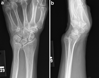
Radiograph of Madelung’s deformity. a posteroanterior view and b lateral view
Fig. 10
Fig. 10
Magnetic resonance images of the ulnar impaction syndrome. a Coronal 2D GRE (TR/TE/flip angle = 533/16/40), b proton-density-weighted (TR/TE = 1531/15), and c STIR (TR/TE/TI = 4389/90/150) images show a bone marrow edema and sclerosis in the lunate (arrows). Diffuse degenerative TFCC tear is noted (arrowhead)
Fig. 11
Fig. 11
Magnetic resonance images of the hamatolunate impingement syndrome. a Coronal 2D GRE image (TR/TE/flip angle = 531/16/40), b proton-density-weighted image (TR/TE = 1,531/15), and c STIR (TR/TE/TI = 4,389/90/150) images show articulation between the hamate and lunate, bone marrow edema, and chondromalacia of these bones (arrows)
Fig. 12
Fig. 12
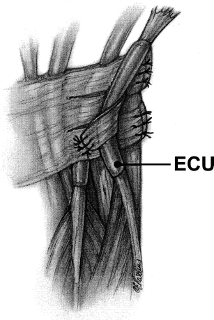
Reconstruction of the extensor carpi ulnaris (ECU) subsheath using adjacent extensor retinaculum. (Reprinted with permission from [152])
Fig. 13
Fig. 13
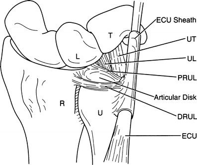
Illustration of the wrist with the TFCC. R radius, U ulna, ECU extensor carpi ulnaris, DRUL dorsal radioulnar ligament, PRUL palmar radioulnar ligament, L lunate, T triquetrum, UL ulnolunate ligament, UT ulnotriquetral ligament. (Reprinted with permission from [153])
Fig. 14
Fig. 14
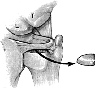
Triangular fibrocartilage complex (TFCC) lesions as described by Palmer. Type I lesions are traumatic in etiology and are subdivided into types IA through ID. a Type IA, central traumatic tear typically located within the sagittal plane within 2 mm from the radial articular surface. b Type IB, medial avulsion lesion that is sometimes associated with an ulnar styloid fracture. c Type IC, distal avulsion lesion with associated ulnocarpal ligament tears. d Type ID, lateral avulsion lesion with tears of the radioulnar ligament and articular disk attachments to the radius. Type ID lesions may have an associated sigmoid notch fracture. e Type II lesion, degenerative in nature and tends to occur in the central avascular region. (Reprinted with permission from [154])
Fig. 15
Fig. 15
Arthroscopic image through a TFCC lesion on the distal aspect of the ulna
Fig. 16
Fig. 16
Ulnar shortening osteotomy. This procedure is performed for positive ulnar variance to relieve ulnar impaction. This can be accomplished by extra-articular ulnar shortening osteotomy or an arthroscopic intra-articular wafer resection. (Reprinted with permission from [152])
Fig. 17
Fig. 17
Hemiresection arthroplasty of the distal ulna with soft tissue interposition. a The fifth extensor compartment of the distal radioulnar joint (DRUJ) is opened, and a dorsal flap of the extensor retinaculum and dorsal DRUJ capsule is elevated. The radial aspect of the distal ulna is resected obliquely and shaped with a burr. b The ulnar edges of the flap of the extensor retinaculum and dorsal DRUJ capsule are sutured to the volar DRUJ capsule. c Axial depiction of the completed hemiresection arthroplasty. (Reprinted with permission from [152])
Fig. 18
Fig. 18
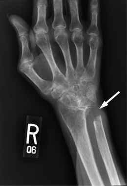
Posteroanterior radiograph of a patient with rheumatoid arthritis treated with the Darrach procedure (distal ulna resection)
Fig. 19
Fig. 19
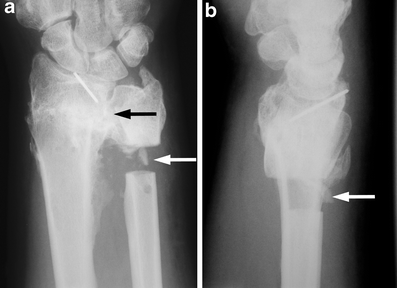
Sauvé–Kapandji procedure. a Posteroanterior and b lateral radiographs of a patient treated with the Sauvé–Kapandji procedure. Black arrow depicts the successful DRUJ fusion. White arrows depict the resected area of the distal ulnar shaft that preserves forearm rotation
Fig. 20
Fig. 20
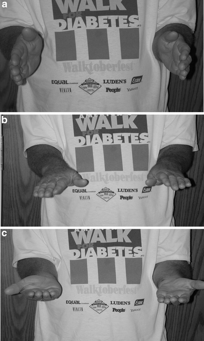
Photographs of a patient treated with the Sauvé–Kapandji procedure demonstrate the preservation of forearm rotation afforded by this operation. Forearm in a neutral rotation, b 90° of pronation, and c 90° of supination
Fig. 21
Fig. 21
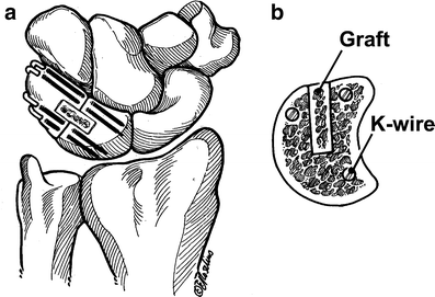
Lunotriquetral (LT) arthrodesis is indicated for LT tears that are unresponsive to arthroscopic debridement. a Posteroanterior plane of LT arthrodesis. b Sagittal oblique plane through the LT arthrodesis site demonstrating correct placement of bone graft and K-wires. (Reprinted with permission from [152])

Similar articles

Cited by

References

    1. Goldfarb CA, Yin Y, Gilula LA, Fisher AJ, Boyer MI. Wrist fractures: what the clinician wants to know. Radiology. 2001;219(1):11–28. - PubMed
    1. Heuck A, Bonel H, Stabler A, Schmitt R. Imaging in sports medicine: hand and wrist. Eur J Radiol. 1997;26(1):2–15. doi: 10.1016/S0720-048X(97)01174-1. - DOI - PubMed
    1. Loredo RA, Sorge DG, Garcia G. Radiographic evaluation of the wrist: a vanishing art. Semin Roentgenol. 2005;40(3):248–289. doi: 10.1053/j.ro.2005.01.014. - DOI - PubMed
    1. Chung KC, Zimmerman NB, Travis MT. Wrist arthrography versus arthroscopy: a comparative study of 150 cases. J Hand Surg [Am] 1996;21(4):591–594. doi: 10.1016/S0363-5023(96)80008-6. - DOI - PubMed
    1. Mikic ZD. Arthrography of the wrist joint. An experimental study. J Bone Joint Surg Am. 1984;66(3):371–378. - PubMed