

Neuropathological heterogeneity in frontotemporal lobar degeneration with TDP-43 proteinopathy: a quantitative study of 94 cases using principal components analysis
- PMID: 20012109
- PMCID: PMC2830004
- DOI: 10.1007/s00702-009-0350-6
Neuropathological heterogeneity in frontotemporal lobar degeneration with TDP-43 proteinopathy: a quantitative study of 94 cases using principal components analysis
Abstract
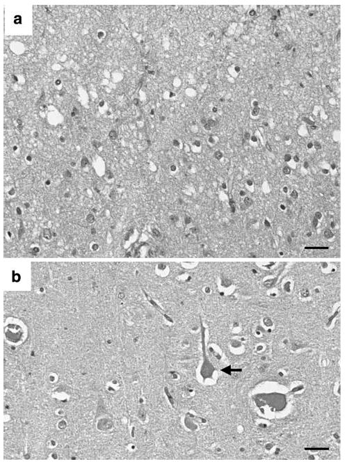
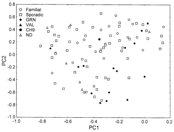
Studies suggest that frontotemporal lobar degeneration with transactive response DNA-binding protein of 43 kDa (TDP-43) proteinopathy (FTLD-TDP) is heterogeneous with division into four or five subtypes. To determine the degree of heterogeneity and the validity of the subtypes, we studied neuropathological variation within the frontal and temporal lobes of 94 cases of FTLD-TDP using quantitative estimates of density and principal components analysis (PCA). A PCA based on the density of TDP-43 immunoreactive neuronal cytoplasmic inclusions, oligodendroglial inclusions, neuronal intranuclear inclusions, and dystrophic neurites, surviving neurons, enlarged neurons, and vacuolation suggested that cases were not segregated into distinct subtypes. Variation in the density of the vacuoles was the greatest source of variation between cases. A PCA based on TDP-43 pathology alone suggested that cases of FTLD-TDP with progranulin (GRN) mutation segregated to some degree. The pathological phenotype of all four subtypes overlapped but subtypes 1 and 4 were the most distinctive. Cases with coexisting motor neuron disease (MND) or hippocampal sclerosis (HS) also appeared to segregate to some extent. We suggest: (1) pathological variation in FTLD-TDP is best described as a 'continuum' without clearly distinct subtypes, (2) vacuolation was the single greatest source of variation and reflects the 'stage' of the disease, and (3) within the FTLD-TDP 'continuum' cases with GRN mutation and with coexisting MND or HS may have a more distinctive pathology.
Conflict of interest statement
Figures


References
-
- Armstrong RA. Correlations between the morphology of diffuse and primitive β-amyloid (Aβ) deposits and the frequency of associated cells in Down's syndrome. Neuropathol Appl Neurobiol. 1996;22:527–530. - PubMed
-
- Armstrong RA. Quantifying the pathology of neurodegenerative disorders: quantitative measurements, sampling strategies and data analysis. Histopathology. 2003;42:521–529. - PubMed
-
- Armstrong RA, Nochlin D, Bird TD. Neuropathological heterogeneity in Alzheimer's disease: a study of 80 cases using principal components analysis. Neuropathology. 2000;20:31–37. - PubMed
-
- Armstrong RA, Lantos PL, Cairns NJ. Spatial correlations between the vacuolation, prion protein deposits, and surviving neurons in the cerebral cortex in sporadic Creutzfeldt–Jakob disease. Neuropathology. 2001;21:266–271. - PubMed
-
- Armstrong RA, Ironside J, Lantos PL, Cairns NJ. A quantitative study of the pathological changes in the cerebellum of 15 cases of variant Creutzfeldt–Jakob disease. Neuropathol Appl Neurobiol. 2009;35:36–45. - PubMed
Publication types
MeSH terms
Substances
Grants and funding
- P50 AG008671/AG/NIA NIH HHS/United States
- P50-AG000658/AG/NIA NIH HHS/United States
- U01-AG16976/AG/NIA NIH HHS/United States
- P50AG008671/AG/NIA NIH HHS/United States
- P50 AG016573/AG/NIA NIH HHS/United States
- P30-AG13854/AG/NIA NIH HHS/United States
- P50 AG005133/AG/NIA NIH HHS/United States
- P50-AG05681/AG/NIA NIH HHS/United States
- P50-AG05133/AG/NIA NIH HHS/United States
- P30 AG013854/AG/NIA NIH HHS/United States
- Z01 AG000658/ImNIH/Intramural NIH HHS/United States
- P01-AG03991/AG/NIA NIH HHS/United States
- P30 NS057105/NS/NINDS NIH HHS/United States
- U01 AG016976/AG/NIA NIH HHS/United States
- P01 AG003991/AG/NIA NIH HHS/United States
- P50 AG005681/AG/NIA NIH HHS/United States
- P50-AG16573/AG/NIA NIH HHS/United States
- K11 AG000658/AG/NIA NIH HHS/United States
LinkOut - more resources
Full Text Sources
Miscellaneous
